



Comparative Pathology Laboratory (Histology)

The University of Delaware’s Comparative Pathology Laboratory focuses on diagnostic surveillance for common and emerging diseases in animal species, including commercial broiler chicken flocks on the Delmarva peninsula, while providing research support for university and regional laboratories.
About | Services | Submitting Specimens | Fees | Getting Results | FAQ | Research Publications | Contact
The Histology/Comparative Pathology Laboratory takes pride in producing high-quality blocks, slides and images through our histology (tissue and slide preparation) services. Additionally, our veterinary anatomic pathologist provides gross and microscopic analysis and interpretation of tissues across organ systems and animal species.
For diagnostics, this lab serves the State of Delaware and Maryland Departments of Agriculture through diagnostic surveillance for disease in Delmarva poultry flocks and other agricultural animal species, giving priority to case submissions from the UD Poultry Health System (Allen Biotechnology Laboratory and Lasher Laboratory) and Maryland Department of Agriculture’s Salisbury and Frederick Animal Health Laboratories.
This service center was established to support the Land Grant, One Health, and Research missions of the University of Delaware, as well as related regional agricultural, animal, and biomedical industries. Our diagnostic surveillance efforts also protect food safety and public health.
Please note: At our current location, we can only receive fixed animal and plant specimens. For whole animal gross evaluation (necropsy), fresh, and unfixed tissues, please arrange with the pathologist for analysis and tissue collection at one of our partner laboratory facilities. Human specimens must be approved by the laboratory Director prior to submission.
This lab provides histology and pathology support for...
- animal health diagnostic laboratories of Delaware and Maryland,
- University of Delaware laboratories and affiliate research groups, and
- external corporations and researchers, upon request.
This section is designed to provide a brief overview of the types of services offered by our laboratory. Please see the accompanying fee schedule for our current service rates.

Gross specimens received in fixative are individually trimmed by an ASCP-certified histotechnician to the size and orientation required for subsequent processing and embedding. Client laboratories also have the option of performing this step prior to submission to reduce preparation fees. The Comparative Pathology Laboratory can provide cassettes and training for proper sample trimming and orientation upon request. Contact us for the current fee schedule.
Trimmed specimens undergo overnight processing in a Leica processor to dehydrate the specimens in preparation for paraffin embedding.
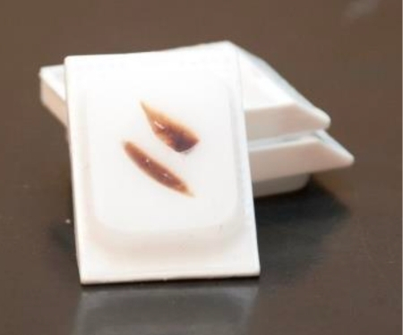
Processed specimens are manually transferred from cassettes to metal wells and are embedded in paraffin wax at a Leica embedding center by the histotechnician to maintain proper specimen orientation for sectioning. Embedded specimens (“blocks”) are complete at this step.
Paraffin blocks are affixed to a Leica microtome and typically cut into 4-6-micron-thick sections. These sections are then transferred from a water bath to a microscope slide for later staining and histopathologic analysis. Unstained slides are complete at this step.
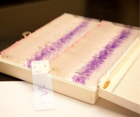
Routine histopathologic analysis utilizes Hematoxylin and Eosin (H&E) staining applied by a Leica autostainer. Special stains are applied manually by the histotechnician. Contact the pathologist for availability and cost. Stained slides are complete at this step.
Bone and other mineralized specimens require chemical decalcification prior to processing to allow for sectioning. This additional step in specimen preparation creates a delay in processing of one or more days until the specimen is deemed “soft” enough to section appropriately. This process can also create loss of detail in soft tissues and bone marrow, which can interfere with microscopic analysis. Please contact the pathologist if this may be a concern in your study.

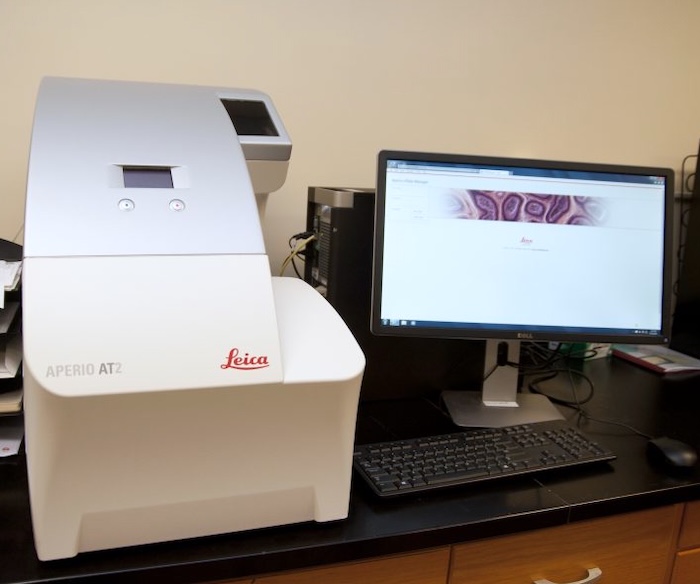
Brightfield or fluorescence scanning of whole prepared glass slides creates permanent virtual slides that can be viewed and analyzed remotely, allowing a personal computer to be used as a high- powered microscope anywhere and anytime an internet connection is available (24/7/365). Slides can be scanned up to a 20x or 40x objective (brightfield) or up to 63x oil (fluorescence). Physical research specimens (tissues, slides and blocks) are returned to the submitting laboratory for storage.
Tissue slides can be analyzed by an ACVP-certified veterinary anatomic pathologist using one of the laboratory's light microscopes or digital pathology systems. Depending upon the needs of the submitting laboratory, the pathologist can provide:
- Description of the lesions observed in the tissues on each slide
- Morphologic diagnosis (concise technical description of the disease process occurring in the tissue)
- Clinical correlation of lesions to signs observed in the animals, to experimental treatments, or to specific disease entities when applicable
- Morphometric analysis (size measurements) and counts of tissue structures and cells
Pathology reports can be delivered by mail, electronically, or in person to a laboratory group and typically include both a written summary and a spreadsheet of pertinent data for each slide, depending on the client laboratory's preference.
High-quality, unaltered digital images of microscopic features of interest are captured by an ACVP-certified veterinary anatomic pathologist and saved in .tif or .jpg format based on client laboratory preference and intended use. Client laboratories have the option of receiving images with or without descriptions and annotations prepared by the pathologist.

Laboratories utilizing the CANR Comparative Pathology Service have unique access to one-on-one or small group consultation and training by a registered histotechnician and/or board-certified veterinary anatomic pathologist prior to or during use of our services.
Consultation and training topics include, but are not limited to: gross specimen collection and submission, specimen trimming and embedding, special stain options, and experimental design related to staining and histologic analysis.
Typically, a brief consultation will be conducted at no charge prior to sample submission to determine individual laboratory needs for services, training, or additional consultation. Sessions can be conducted by email, phone, or in person based upon client preference.
Training can be conducted in our laboratory (for trimming and embedding) or in the client’s laboratory (for trimming) based upon client preference. Following training, clients may also elect to self-embed tissues using our equipment during normal business hours to reduce the cost of tissue preparation.
Equipment
In support of specimen preparation, the laboratory has dedicated stations and equipment for:
- Trimming- chemical hood
- Paraffin embedding- Leica EG1150H Embedding Center
- Tissue processing- Leica ASP300 Tissue Processor
- Microtomy- Leica RM 2255 Automated Microtome
- Automated slide staining- Leica AutoStainer XL ST5010
Laboratory clients are invited to use the Olympus BX40 microscope with Nikon DS-Fi2 camera and corresponding NIS Elements image analysis software for analysis.
General Requirements
Specimens should NOT be frozen or shipped with frozen materials to prevent irreversible tissue damage that could impair microscopic analysis.
Specimens must be received in...
- clean leak-proof specimen containers with adequate fixative (10:1 fixative to tissue ratio by volume),
- clearly labeled with specimen ID information and fixative hazard labels, and
- triple-packaged (see below) to prevent chemical leakage and exposure during transit or specimen accessioning.
Visibly dirty (i.e., blood or feces-coated), unlabeled, improperly labeled, or leaking containers may be rejected and/or, in egregious cases, result in federal fines to the submitting party.
Triple packaging
Assure primary container is tightly sealed. Secure the cap with tape or parafilm.
Wrap primary container in sufficient absorbent material to absorb the entire fixative amount if the container were to break or leak.
Place the primary container and absorbent into a secondary container such as another larger screw-top container or a heavy duty ziplock bag.
Use a sturdy shipping box in good condition. Add padding or styrofoam as needed to reduce specimen movement.
Include a piece of paper inside the box stating that the samples are not regulated per DOT or IATA. Describe the samples on this sheet or on a laboratory accession form.
Specimens can be shipped via FedEx/UPS or transported in designated university vehicles to the laboratory:
Address
Delaware Biotechnology Institute, Ste. 140
15 Innovation Way
Newark, DE 19711
Hours
8 a.m. to 5 p.m. Monday through Friday; The laboratory building is closed and cannot receive shipments on university-recognized holidays.
Phone
302-831-7208
- Cut the tissue sample using clean decisive strokes with a sharp scalpel blade, razor blade, knife or scissors—sawing motion with a blade damages tissues.
- Specimens should be < 0.5 cm thick in at least one plane. If in cassettes, tissue should not touch the walls or lid of the cassettes for proper fixation.
- Minimize tissue handling with forceps (especially rat-toothed forceps) and fingers, which can distort histologic features. Likewise, do not scrape mucosal surfaces (e.g., the intestine or trachea) with blades or fingers to remove debris.
- Immerse the sample directly into the fixative to prevent desiccation. Maintain a 10:1 ratio of fixative to tissue volume for proper fixation.
- Label specimen containers with unique specimen IDs, the tissues/organs they contain, and the type of fixative. If submitting tissues in cassettes, each cassette should be labeled with a unique specimen ID number using a pencil on a flat side. Inks, including permanent marker, are removed by the alcohols used in tissue processing and the front (“angled”) side of the cassette is utilized by the histotechnician to record the pathology accession number for future reference.
- Containers with formalin must have formalin-hazard labels affixed.
- Specimen containers must be leak-proof, clean, and dry.
- Submit a completed accession form with the specimens. If histopathologic analysis is desired, please provide thorough information regarding study design, the experimental groups and treatment course, and clinical signs and gross lesions in animals and tissues. The Comparative Pathology Laboratory “FAQ” provides additional details.
- Please drop off specimens at the laboratory during normal business hours. If no one is available in the laboratory, specimens can be left with staff at the Department of Animal and Food Sciences Office in room 044, Townsend Hall (immediately across the sidewalk from Worrilow Hall).
Fee Schedule
Effective July 1, 2026. Contact us with any additional questions.
Histology Services (Tissue block and slide preparation)
If microscopic analysis and interpretation of slides is also required, please see pathology service fees below.
Trimmed tissue submitted in cassette
| Service | Cost ($) per cassette unless noted |
|---|---|
| Process only (Self-embed) | 4 |
| Process, Embed (Block only) | 5.50 |
| Process, Embed, 1 unstained slide | 7.50 |
| Process, Embed, 1 H&E slide | 9.50 |
Untrimmed tissue submitted in fixative
| Service | Cost ($) per cassette unless noted |
|---|---|
| Trim, Process, Embed (Block only) | 8.50 |
| Trim, Process, Embed, 1 unstained slide | 10.50 |
| Trim, Process, Embed, 1 H&E slide | 12.50 |
Recut slide from prepared block
| Service | Cost ($) per cassette unless noted |
|---|---|
| Additional unstained slide | 4.25/slide |
| Additional H&E slide | 5.25/slide |
Additional histotechnician support
| Service | Cost ($) per cassette unless noted |
|---|---|
| Consultation/Training in trimming & embedding | 75/hour |
| Decalcification of bone/mineralized specimen | 5/specimen or block |
| Special stains (PAS, Gram, GMS, etc.) | Contact pathologist |
| External diagnostic testing of archived specimens | Direct test cost + S&H |
Pathology Services- Pathologist Consultation, Gross & Microscopic tissue analysis
Requires prepared slides, see above for tissue preparation fees.
| Service | Cost ($) per cassette unless noted |
|---|---|
| Histopathologic analysis of stained slides | 7.50/slide + Reporting Fee below |
| Pathologist Consultation and Reporting Fee | 150/hour* |
*NOTE: Please contact the pathologist for an estimate of time required based upon the complexity and number of specimens for the analysis. The pathologist will assist researchers with preparation of related grant proposals and publications at no additional cost.
Services in this category are based upon the needs of the submitting laboratory and include:
- Research design consultation
- Analytic reporting and data spreadsheet preparation from gross or microscopic analysis
- Student training in anatomic tissue localization and collection
- Animal autopsy (necropsy) with gross lesion detection and interpretation
- Digital image capture with description/annotation
Processing Fees
| Service | Cost ($) per cassette unless noted |
|---|---|
| In network (UD affiliate) orders | N/A |
| Surcharge for Rush and Out-of-network (non-UD affiliate) orders | +25% of total order |
Quality Assurance
We take great pride in producing high-quality blocks, slides and images through our service. Please contact the pathologist with any concerns about specimen or image quality. We will make every effort to resolve quality control issues promptly and will replace slides with artifacts related to trimming, sectioning or staining at no cost to the client if these services were performed in our laboratory.
Expected turnaround time
Originally developed as a support laboratory dedicated to the surveillance and diagnosis of disease in Delmarva poultry flocks, our laboratory gives the highest priority to diagnostic case submissions from the UD Poultry Health System (Lasher Laboratory and Allen Laboratory) and from Maryland Department of Agriculture’s Salisbury and Frederick Animal Health Laboratories. Diagnostic case results are reported within 1-5 business days.
Our laboratory has a single histotechnician and veterinary pathologist. Research studies have a targeted completion of one to three weeks for histology services and two weeks to two months for histopathologic analysis, depending on the complexity of analysis and specimen numbers. Please contact us for an estimate of the time to completion for your submission, as diagnostic and research caseload is highly variable throughout the year. Rush orders are available for an additional fee.
Retrieving finished materials (blocks or slides)
Research specimens and tissue blocks, and slides produced from research specimens are considered the property of the submitting client and are returned upon completion of the work request. Due to space limitations, we can no longer add research specimens to our archives, so submitting laboratories will be notified to claim materials at study completion. The laboratory reserves the right to permanently dispose of research specimens not claimed within 6 months of study completion.
Diagnostic case materials are the property of the Comparative Pathology Laboratory and are archived in our specimen library in Suite 140 at 15 Innovation Way and off-site storage. Blocks and slides in the diagnostic specimen archive are retained for approximately 20 years. Archived material can be utilized for teaching or research upon request, but must be returned to our laboratory following use.
Please note
Once research materials (tissues/blocks/slides) are removed from the Comparative Pathology Laboratory, it is the responsibility of the client to properly store and maintain the materials. The Comparative Pathology Laboratory cannot be held responsible for lost, broken, or damaged specimens following removal from our laboratory. Recuts of lost or broken slides can be made for an additional fee if original blocks are still available.
We do not currently have the equipment to produce frozen sections, though we are considering the addition of this service in the near future. In the meantime, cryostat sectioning is available through the Delaware Biotechnology Institute (DBI) Imaging Core Facility and the A.I. duPont Children’s Hospital Histology Laboratory.
At present, our laboratory only performs special stains for disease organisms commonly encountered in diagnostic service (Gram stain, GMS, etc.). We can advise laboratories on other stains that may be applicable for a specific area of interest and can order and perform these additional staining procedures upon request. Please contact the pathologist for more information.
We welcome the submission of fixative containers with either pre-trimmed tissues in cassettes or untrimmed tissues. Feel free to contact the pathologist if you require assistance with tissue collection and handling or proper orientation in cassettes.
Trimming your own tissues is also a way to save on service fees, and our laboratory can provide cassettes at a nominal cost. However, tissues do not have to be submitted in cassettes, and our histotechnician routinely trims specimens submitted to our service.
Specimens must be submitted in an adequate volume of fixative regardless of whether they are whole or pre-trimmed in a cassette. Containers with formalin must have appropriate formalin-hazard labels. For further information, please review our sample submission guidelines.
We can also train users on proper embedding techniques upon request. Trained users can then self-embed tissues at our embedding station upon arrangement with the histotechnician. Due to liability issues and potential costs related to service and repair following non-technician use, we cannot currently allow clients to perform microtomy (sectioning) using our equipment.
Just as you would not want your doctor to attempt diagnosis of an illness without hearing about your symptoms and performing a physical examination, the pathologist needs to be informed of important clinical and specimen information before microscopic analysis.
This information includes:
- Animal information: Species, breed, sex and castration status, age
- Clinical information: Duration and signs of disease, results of other diagnostic testing
- Specimen information: The organ or tissue and specific anatomic location of each specimen, how the specimen was collected, and gross lesions observed in the specimen. For lesions, please describe: size (measure if possible), shape, color, consistency, distribution.
- NOTE: The pathologist typically does not observe the gross specimen prior to tissue processing and sectioning. It is imperative to describe what you saw in the body and specimen prior to trimming and fixation.
- Clinical reasoning: What insight are you hoping to gain from microscopic tissue analysis? What lesions are you expecting the pathologist observe (for experimental disease agent exposures)? What is/are your working diagnosis or differential diagnoses?
- Research protocol: Experimental groups, known disease exposure, group treatments and time course, etc. (for research cases only). Additional protocol sheets can be attached to the submission form as needed.
A summary of the information listed above should be provided with the specimen accession form. Printed protocols and other supplemental information (digital photos, diagnostic test reports, etc.) can be attached to the accession form if necessary.
Please review the section titled Expected turn-around time, outlined above.
Research Publications
Expectations of Acknowledgement or Authorship
Since our role in research involves generating a portion or all of the histologic specimens (blocks/slides) and/or histopathologic analysis for a given study, it is important for client laboratories to acknowledge the UD Comparative Pathology Laboratory or its staff in presentations and publications that result from our work. While authorship is not required for work performed in our laboratory, inclusion of a certified pathologist or histotechnician may be beneficial for the purposes of manuscript preparation and peer-review. Please consider authorship for the pathologist or histotechnician when our staff is significantly involved in experimental design and sample preparation, in generating a major portion of results and analysis, and/or in grant proposal and manuscript preparation. We thank you in advance for acknowledging our efforts and contributing to the future success of our laboratory!
Support for Writing for Grants or Publication
We are happy to contribute to collaborative grant applications or publications originating from work performed by our laboratory upon request. This includes, but is not limited to, writing or consulting on budget preparation and sections related to materials and methods, results, analysis, or discussion involving histology and histopathology.
Recent Publications and Grants
Content arising from work performed in the University of Delaware Comparative Pathology Laboratory.
Publications
- Michael B. Papah, Erin M. Brannick, Carl J. Schmidt, Behnam Abasht, Arda Yildirim. Gene expression profiling of the early pathogenesis of wooden breast disease in commercial broiler chickens using RNA-sequencing. PLOS ONE 13:12, pages e0207346, 2018.
- Michael P. Babak, Erin M. Brannick, Carl J. Schmidt, Behnam Abasht. Evidence and Role of Phlebitis and Lipid Infiltration in the Onset and Pathogenesis of Wooden Breast Disease in Modern Broiler Chickens. Avian Pathology, 2017.
- Jack Gelb, Jr., Daral J. Jackwood, Erin M. Brannick and Brian Stewart Ladman. Efficacy of Recombinant HVT-IBD Vaccines Administered to Broiler Chicks from a Single Breeder Flock at 30 and 60 Weeks of Age. Avian Diseases, 60: 3, 603-612, 2016.
- Marie F. Mutryn, Erin M. Brannick, Weixuan Fu, William R. Lee and Behnam Abasht. Characterization of a novel chicken muscle disorder through differential gene expression and pathway analysis using RNA-sequencing. BMC Genomics, 16: 399, 2015.
- Rogers, A., E. Pritchett, R. Alphin, E. Brannick, and E. Benson. I. Evaluation of the impact of alternative light technology on male broiler chicken growth, feed conversion, and allometric characteristics. Poultry Science, 94, 408-414, 2015.
- Rogers, A., E. Pritchett, R. Alphin, E. Brannick, and E. Benson. II. Evaluation of the impact of alternative light technology on male broiler chicken stress. Poultry Science, 94, 331–337, 2015.
- Gelb, J. Jr., B. S. Ladman, C. R. Pope, J. M. Ruano, E. M. Brannick, D. A. Bautista, C. M. Coughlin, and L. A. Preskenis. Characterization of Nephropathogenic Infectious Bronchitis Virus DMV/ 1639/11 recovered from Delmarva broiler chickens in 2011. Avian Diseases, 57: 1, 65-70, 2013.
Grants
- Dyer, R.M., T.F. Gressley, R. Arsenault, and E. Brannick. 5/16-6/17. UD CANR Seed Grant. $50,000. Crosslinkage between gut fermentation, adiposity and immune response activates mesenteric adipose depot inflammation in dairy cows.
- Gelb, J. Jr., B. S. Ladman, and E. M. Brannick. (USPEA F065). 7/1/15-6/30/17. $71,979. Evaluation of Cross-Protection Provided by IBV Vaccination Programs vs. Challenge with Current U.S. Variant Viruses. U.S. Poultry & Egg Association (USPEA).
- Abasht, B. et al. USDA-NIFA. $500,000. 3/1/16-2/29/20. Genome-Wide Identification and Functional Validation of Genes Causing Susceptibility To Wooden Breast Disease In Commercial Broiler Chickens.
- Gelb, J. Jr., B. Ladman, and E. Brannick. $71,979. 7/1/15-6/30/17. U.S. Poultry & Egg Association. Protection provided by multivalent infectious bronchitis virus vaccines against challenge with current U.S. strains and variants.
- Brannick, E.M. and J. Gelb, Jr. UNIDEL. 02/15-01/16. $550,100 Virtual Microscopy- Collaborative Teaching, Research, & Outreach.
- Abasht, B., E. Brannick, and C. Schmidt. U.S. Poultry & Egg Association (USPEA F059). 6/14-5/16. $57,919. Identifying the Onset of a Novel Muscle Disorder in Chickens through Differential Gene Expression and Histologic Analyses.
- Gelb, J. Jr., B. Ladman. and E. Brannick. U.S. Poultry & Egg Association (USPEA
11A00887). 8/11-7/13, extension through 2/14. $70,734. Studies on the Efficacy of Recombinant HVT-IBD Vaccines. - Gelb, J. Jr., B. Ladman, and E. Brannick. U.S. Department of Agriculture (USDA AI CAP Subaward Z541310, with University of Maryland). 2/11-1/13. $86,406. Viral Adaptation to Host Species-Effect of AIV Passage within a Host on Viral Populations and Sequence Changes FY11.
Hours
8 a.m. to 5 p.m. Monday through Friday
Phone
302-831-7208

Erin Brannick, DVM, MS, ACVP Diplomate
Director of Undergraduate Programs
Office Location:
531 S. College Avenue
041 Townsend Hall
Newark, DE 19716
Dr. Brannick is broadly trained in veterinary medicine and pathology through the Ohio State University College of Veterinary Medicine. She is board-certified in veterinary anatomic pathology by the American College of Veterinary Pathologists (ACVP) and serves as an Associate Editors for Zachary’s Pathologic Basis of Veterinary Disease, one of the premier training texts for veterinarians and veterinary pathologists worldwide. She has been in the field of veterinary pathology since 2010 and has over three decades of veterinary experience.

Joanne Cooper
Office Location:
Delaware Biotechnology Institute
Suite 140
15 Innovation Way
Newark, DE 19711
Joanne Cooper is a registered histotechnician certified by the American Society for Clinical Pathology (ASCP). She received her training from Delaware Technical Community College (DTCC) and has almost two decades of histology experience.
She helps to train future histotechnicians in the UD Comparative Pathology Laboratory through a training partnership with DTCC and is active in the Delaware chapter of the American Society for Clinical Laboratory Science.