
![CAS-CHEM-Booksh-Raman-AFM-072619 Karl Booksh, professor of Chemistry and Biochemistry, has been a campion of UD’s acquisition of a new atomic force-Raman microscope. He and his team focus on research into chemometrics, spectroscopy, and plasmonics and “in addition to revealing chemical information about a sample, the microscope provides much higher resolution than available previously about the surface of a material, its electrical and thermal properties.“ [UDaily]](/udaily/2019/october/atomic-force-Raman-microscope-Karl-Booksh/_jcr_content/udaily_Image.coreimg.jpeg/1570038919127/11-karl-booksh-raman-microscope-cas-chem-afm-072619-ek011-800x533.jpeg)
Scoping out new science
Photos by Evan Krape; microscopic images courtesy of Booksh Lab Group October 02, 2019
New microscope with dual capabilities supports multitude of studies
A single strand of DNA. The toxic pollutants in a waft of air. A paint sample from a priceless work of art. Flakes of a Martian meteorite. That’s only a smattering of what scientists will be able to examine with the new microscope — an atomic force-Raman microscope, to be exact — now housed in the University of Delaware’s Lammot du Pont Laboratory.
“UD is excited to add this important and state-of-the-art new tool to our suite of instruments for examining materials at high resolution,” said Charles G. Riordan, vice president for research, scholarship and innovation. “With this capability, UD faculty, students and staff will be able to drive research and education forward in a wide array of fields, from engineering to physical sciences to art conservation.”
The new microscope will help researchers go where they couldn’t before. Previous scopes just didn’t have the super-high resolution and chemistry-uncovering power this one has.
“This microscope will allow scientists to see objects 10,000 times smaller than the diameter of a human hair — plus provide detailed information about both the surface of a material and its chemistry,” said Karl Booksh, professor of chemistry and biochemistry and the rallying force behind UD’s successful proposal to the National Science Foundation. NSF came through with a $558,228 grant from its Major Research Instrumentation and Chemistry Research Instrumentation programs and the Established Program to Stimulate Competitive Research (EPSCoR). The UD Research Office also helped support the cost of the instrument, which was purchased from Horiba, a leading provider of analytical and scientific measurement systems.
![CAS-CHEM-Booksh-Raman-AFM-072619 Karl Booksh, professor of Chemistry and Biochemistry, has been a campion of UD’s acquisition of a new atomic force-Raman microscope. He and his team focus on research into chemometrics, spectroscopy, and plasmonics and “in addition to revealing chemical information about a sample, the microscope provides much higher resolution than available previously about the surface of a material, its electrical and thermal properties.“ [UDaily]](/udaily/2019/october/atomic-force-Raman-microscope-Karl-Booksh/_jcr_content/par_col_8_udel/image.coreimg.jpeg/1570039075405/22-karl-booksh-microscope-cas-chem-raman-afm-072619-022-800x533.jpeg)
This new tool is a “scientific twofer,” combining two microscopes in one. A Raman microscope, named after the late Indian physicist and Nobelist Sir Chandrashekhara Venkata Raman, scans a sample with a laser, interacting with the vibrations of the molecule of interest, scattering the light. These light patterns serve as “fingerprints” for identifying the molecules and for studying their chemical bonds and degree of interactivity with other molecules.
An atomic force microscope scans a sample using a small probe that yields information about the surface, such as its topography, hardness, electrical and thermal properties. This probe, tipped in gold, is nearly “atomically sharp,” meaning it is virtually able to detect a single atom.
Combining both techniques within a single microscope delivers a trove of information simultaneously. And that’s important for a number of studies across the University and with industry collaborators, as well as partnering institutions such as Winterthur Museum.
Putting the scope to work
During the summer of 2019, doctoral student Devon Haugh and undergraduate Savannah Talledo, a Wofford College student participating in the NSF-funded Science and Engineering Leadership Initiative at UD, used the new microscope to study air pollutants. Tiny gas particles from vehicle exhaust and soot generated from burning coal can fuel climate change and increase the risk of asthma, lung disease, heart disease and other health problems. The microscope helped to determine the acidity of the airborne particles, which influences how quickly they will grow in the atmosphere.
“Understanding acidity can help us improve predictions of how airborne particles affect human health and climate,” said Murray Johnson, professor of chemistry and biochemistry, who is leading the project. “In a conventional laboratory, acidity is measured with a pH meter. However, that approach does not work for airborne particles on the sub-micrometer-size scale, hence the need for new measurement approaches such as the Raman microprobe.”

Haugh was glad to have access to the new instrument for her work.
“I care about the health of our environment,” she said. “This project allows me to contribute toward better understanding and protecting it.”
Experts at Winterthur’s Scientific Research and Analysis Lab will focus the microscope on the museum’s valuable collections of historic textiles, as well as its Chinese export paintings from the 18th and 19th centuries, according to Jocelyn Alcántara-García, assistant professor and a co-investigator on the grant. In the first half of the 19th century, with the boost in foreign trade due to the opening of ports in China, a large number of Western synthetic chemical pigments were imported to China. Before long, these man-made pigments replaced the mineral and plant pigments that Chinese painters had traditionally used in their artwork, from watercolors to reverse-painted glass. The new microscope will help conservation scientists gain a better understanding of this transitional period.
Alcántara-García said she will use the instrument to understand the fixatives that were used to set the dye in historic textiles, which will help textile conservators and other museum professionals determine degradation mechanisms and potential interventions.
Solving challenges on Earth and Mars
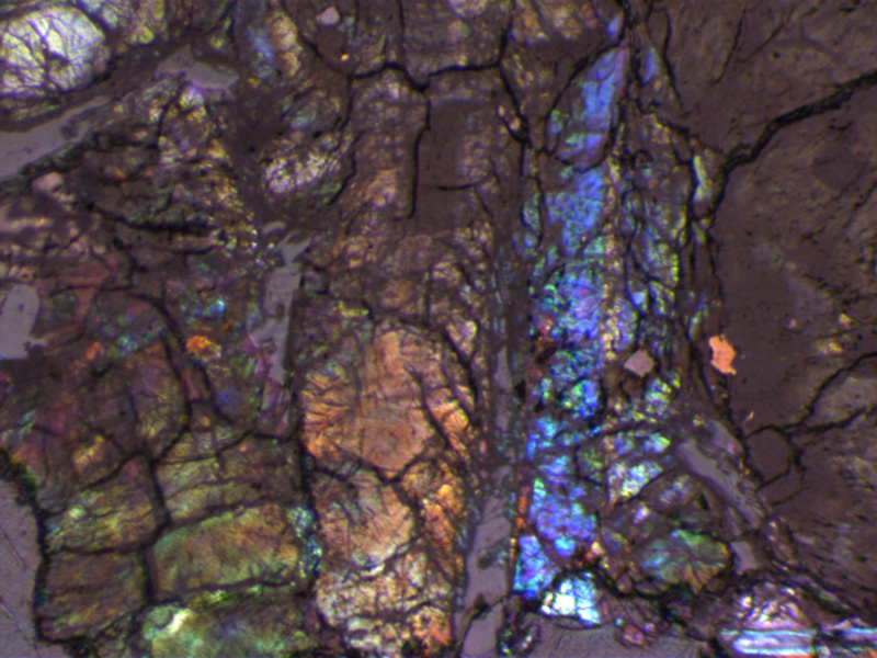
Now, about those meteorites … in a collaboration that began when he joined the UD faculty a decade ago, Booksh is working with Merck senior scientist Joseph P. Smith, who earned his doctorate in analytical chemistry at UD, and with Marietta College professor Frank Smith, who earned his doctorate in geology at UD, to unlock some of the secrets of the planets through clues provided by lunar, Martian and asteroidal meteorites. The samples came to the team on good authority — from NASA’s Johnson Space Center and from the Smithsonian.

The team’s primary interest is the chemical composition and properties of these rocks, which contain “shock pockets” created from all the fracturing and melting that occurred when they hit the ground. Their chemistry can help reveal the geology and atmospheres of their home planets. Smith said the work also could aid the search for life on Mars in NASA’s and the European Space Agency’s 2020 rover missions.
“The NASA and ESA rovers both will have, for the first time, Raman spectrometers to help characterize Martian surface materials,” Smith said. “As such, our work investigating meteorites may help enhance the search for life on Mars by developing optimal data collection and analysis methodologies.”
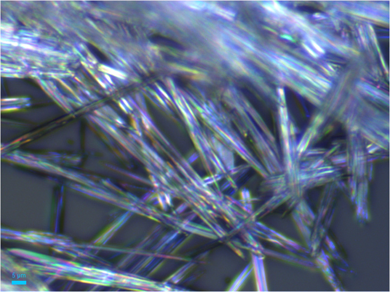
Booksh and Smith also are working on other intriguing problems right here on Earth—as collaborators on Merck & Co. Inc. and UD project focusing on pharmaceutical applications. The team will investigate polymorphism in drug development — the ability of a solid to exist in two or more crystalline forms, each with vastly different physical and chemical properties. Polymorphs are of particular concern to the drug industry because one of these forms may be toxic, and more than 50 percent of active pharmaceutical ingredients have more than one polymorph.
“We’re hoping to develop the next generation of analytical techniques that will help solve these complex challenges facing the pharmaceutical industry,” Smith said.
Contact Us
Have a UDaily story idea?
Contact us at ocm@udel.edu
Members of the press
Contact us at 302-831-NEWS or visit the Media Relations website

