




Plant biology breakthrough
Photos by Kathy F. Atkinson and courtesy of the Caplan Lab October 13, 2026
UD-developed method helps researchers study proteins involved in plant health, disease
Plants — like people — come in all shapes and sizes.
And just like scientists want to understand the components of the human body, to treat disease and improve our lives, they also want to understand how plants thrive, get disease, succumb or survive.
It’s a real problem that affects us, too. One powerful way scientists investigate these questions is through proteomics, the study of the proteins that live and work inside cells. This is true whether those cells reside inside a human body, an animal, a plant or a virus.
While much ground has been gained in understanding human biology using proteomics, progress has been slower for those studying plants.
This is because plant tissues (think leaves, pollen, seeds) have a hard exterior shell and a thick cell wall to protect the proteins and other essential components inside and to keep unwanted materials out. These protections serve a purpose but also create a barrier, scientifically speaking.
Now, University of Delaware researchers report in the journal Plant Physiology a validated method to digest proteins inside of intact plant cells, overcoming this long-standing hurdle in plant proteomics.
Yanbao Yu, director of proteomics in UD’s Department of Chemistry and Biochemistry, said it represents a “breakthrough for the plant proteomics community” that opens a new window for plant biology to answer previously difficult-to-study questions.
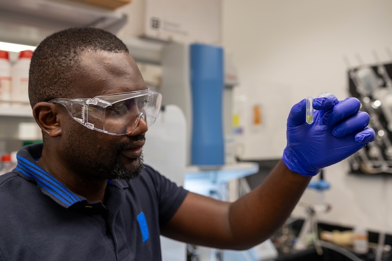
For example, the ability to detect significant responding proteins could provide a biomarker to indicate that a plant has a particular bacterial or fungal infection, Yu said, or potentially lead to ways to deliver proteins as a therapeutic to help cells recover from disease.
He hopes the approach can inform the agricultural community’s bottom line.
“Plant infection is an economically important problem for agriculture,” said Yu. “With this new method to study plant infection, we can identify biologically meaningful infection-related proteins that can improve agriculture.” He previously demonstrated the UD-developed technology’s effectiveness in simple cell lines.
How does in-cell digestion work?
Traditionally, researchers studying plant proteins must first grind up the tissue using a mortar and pestle and put it through a series of chemical processing steps to isolate the proteins they want to study. The process can take days, and valuable material can be lost along the way.
Yu’s method takes a different approach. Rather than extracting the proteins first, researchers treat the intact plant tissue with methanol, which makes it possible to study the proteins inside the cells while preserving important information.
“It acts like a blast freezer,” said Yu. “It freezes the biology inside the cell so that it can be studied.”
In essence, the methanol breaks the cell open, but the original pathology inside the cell remains unchanged. From these broken cells, Yu said, it’s possible to cut, remove and quantify the exact proteins inside.
Methanol fixation is common in pathology and other imaging analysis, used to keep the tissue structure intact, but few researchers have used it for proteomics analysis, because they generally want to break the cells apart and work with proteins directly.
By contrast, Yu decided to digest proteins while the cells are still intact.
“We don't destroy it using a missile; we use a chemical like a knife to cut inside,” he said.
And because the UD-developed proteomics method is so sensitive and robust, it is possible to accurately identify large numbers of proteins consistently, even from a limited amount of sample material.
Understanding fungal infection in tobacco leaves
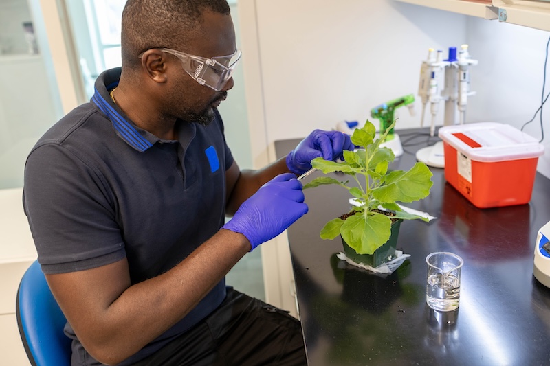
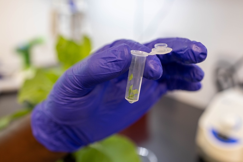
Collaborating with Jeff Caplan, associate professor of plant and soil sciences and director of the Delaware Biotechnology Institute’s Bio-Imaging Center, and Deji Adekanye, a postdoctoral researcher in the Caplan lab, Yu applied this strategy to investigate healthy and fungal-infected tobacco leaves to learn how the plant’s immune system responds to infection.
They wanted to understand what makes a fungus a pathogen, since fungi are all around us, but only cause disease on a limited number of plant species. To tease this apart, Yu investigated how a plant’s protein profile changes in response to disease-causing and harmless fungi. The data showed distinct differences in the proteins present.
“In terms of biology, we identified a lot of signature proteins that people just wouldn’t be able to see with traditional methods,” said Yu. “And we can clearly see the protein response.”
The research team’s analysis showed that plant extracellular vesicles responsible for cell-to-cell communication were enriched with proteins involved in immune functions.
“This suggests an active plant defense role for these vesicles,” said Adekanye, the paper’s first author.
Meanwhile, the data showed extracellular vesicles from the fungal pathogen were full of poisonous proteins that can attach to a protein or enzyme inside a cell and act like a switch, turning on infection. The pathways plants use to respond to disease-causing and harmless fungi were different, too.
It's valuable information to have, Adekanye said, as he continues to explore the role of extracellular vesicles in plant-fungal interactions.
The method is not limited to plants, either, showing efficacy in many other biological systems and model organisms, including Caenorhabditis elegans worms, frog embryos, sea urchin, bacterial cells, neuroblastoma cells and yeasts. To date, faculty and students from over 15 laboratories on campus have leveraged Yu’s UD-developed technology platform, which is licensed by CDS Analytical, LLC, a leading supplier of proteomics sample preparation consumables and instruments, to study a wide range of questions in developmental biology, infection and immunology, chronic nerve pain and more.
“I believe this is going to be a game changer. It’s really a practical yet powerful way of doing protein sample preparation with small number of cells, and potentially single cells,” Yu said.
Additional coauthors on the paper include 2026 UD alumnae Jasmine Parks, who earned a bachelor’s degree in English and in biochemistry, and Meghara Kusuru, who earned a master’s degree in bioinformatics and computational biology.
Contact Us
Have a UDaily story idea?
Contact us at ocm@udel.edu
Members of the press
Contact us at mediarelations@udel.edu or visit the Media Relations website