

Why Achilles tendon injuries resist treatment
Photos by Kathy F. Atkinson November 07, 2025
UD researchers seek to uncover how anatomical differences may influence recovery
For people with pain in the Achilles tendon, the band of tissue connecting the calf muscles to the heel, recovery can be slow and unpredictable. Even with the best available therapy, many continue to struggle with recurring pain that can linger for years.
Achilles tendinopathy refers to injuries that result in pain and stiffness in the tendon, causing difficulty with movement. The condition can arise when the tendon is strained from repeated overuse during physical activity and is associated with factors like aging, metabolic disorders or obesity.
The gold-standard treatment for Achilles tendinopathy is a progressive series of calf-strengthening exercises. Yet even with this therapy, up to 60% of patients still report pain five years after diagnosis.
“The complex anatomy of the Achilles tendon varies across individuals, which may contribute to the inconsistent results of the current one-size-fits-all treatment approach,” said Stephanie Cone, assistant professor at the University of Delaware’s College of Engineering. “We seek to better understand how anatomy relates to Achilles tendinopathy injuries so we can develop more effective treatments in the future.”

A $3.2 million, five-year R01 grant from the National Institutes of Health will support the work. Cone's laboratory in the Department of Biomedical Engineering is partnering with researchers at UD’s College of Health Sciences and the University of North Carolina at Chapel Hill.
“This is a truly interdisciplinary effort, bringing together engineers, clinicians, imaging experts and computational modelers,” said Cone.
Understanding responses to exercise treatment
The Achilles tendon comprises three bundled subtendons, each linked to one of three calf muscles: two gastrocnemius and one soleus. “Basically, you’ve got three motors—the muscles—that are pulling on three cords wrapped around each other—the subtendons,” explained Cone.
Past work with healthy volunteers revealed that this anatomy varies from person to person. Some people have a larger soleus subtendon, while others have a gastrocnemius subtendon that dominates. The new study will explore how these anatomic variations affect Achilles tendinopathy injuries and recovery.

The researchers suspect that the standard exercises benefit some patients because they happen to engage the subtendon most affected by injury. Others may see less benefit because the exercises do not directly target the right region.
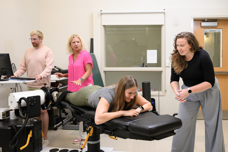
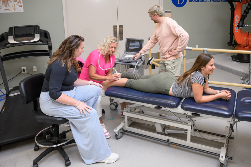
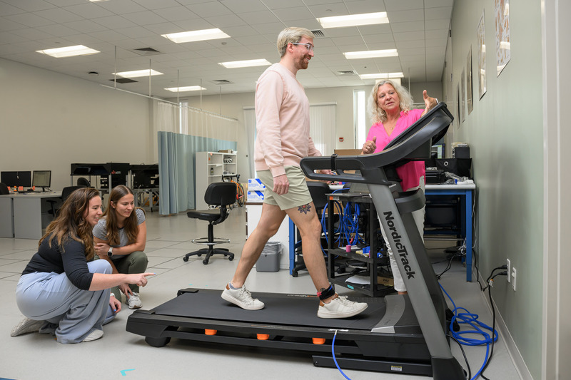
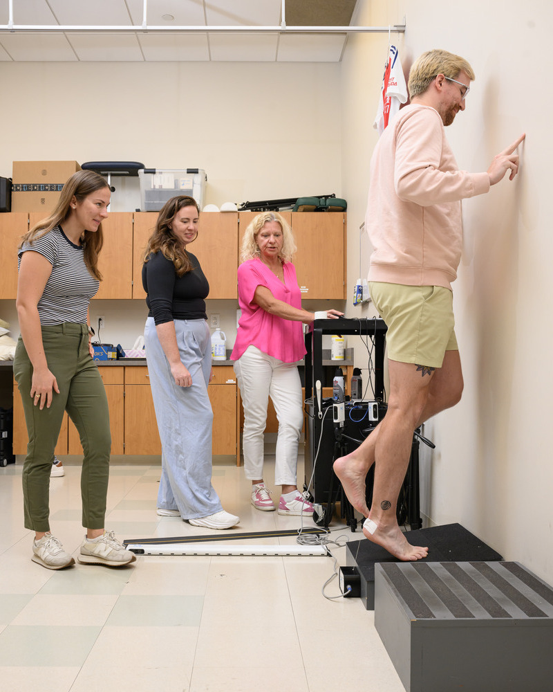
Part of the work will be performed in collaboration with Karin Grävare Silbernagel’s team at the Delaware Tendon Research Group in UD’s Department of Physical Therapy. Study volunteers will receive standard 12-week treatment for Achilles tendinopathy provided by a physical therapist. They will complete movement tests and MRI scans before and after to track improvements in muscle strength and tendon structure and function.
In general, most patients with Achilles tendinopathy experience noticeable improvement after 12 weeks of physical therapy, although recovery is not complete and progress often continues at home. Physical therapists typically recommend continuing the exercises even after pain subsides.

“We see how frustrating Achilles tendon injuries can be. You do the exercises, feel better, stop and the pain comes back. If we can find ways to break that cycle or tailor what works best for each person, it would make a big difference,” said study investigator Andy Smith, a physical therapist and doctoral candidate in UD’s Biomechanics and Movement Sciences Program.
The UD team plans to recruit 50 volunteers with Achilles tendinopathy, with study enrollment expected to begin in November.
Zooming in on anatomical differences
A second part of the project leverages Cone’s long-standing connections with UNC, where she completed her doctoral research. UNC’s imaging center houses a 7-Tesla MRI scanner capable of recording images at much higher resolution and contrast than standard MRI.
The North Carolina team, led by Jason Franz, will recruit local participants with Achilles tendinopathy to receive these ultra-high-resolution MRI scans. The results will give the researchers a detailed, three-dimensional view of individual injuries within the Achilles subtendons.
Franz, a professor in the Lampe Joint Department of Biomedical Engineering at UNC and North Carolina State University, will collaborate with Geoffrey Handsfield, assistant professor of orthopaedics and biomedical engineering at UNC.
The researchers will use these 3D scans to create computational models of Achilles tendon injuries. These models will allow the researchers to explore how specific exercises stimulate the injured muscle and activate the affected subtendon.
“We hope our research will pave the way for future development of more personalized approaches to treating Achilles tendinopathy,” said Cone.
Funding is provided by NIH’s National Institute of Arthritis and Musculoskeletal and Skin Diseases under award number AR085639.
Contact Us
Have a UDaily story idea?
Contact us at ocm@udel.edu
Members of the press
Contact us at mediarelations@udel.edu or visit the Media Relations website

