
Digital pathology
Photo by Wenbo Fan July 26, 2016
UD Comparative Pathology Lab serves campus, regional partners
The University of Delaware’s Comparative Pathology Laboratory, housed in the College of Agriculture and Natural Resources (CANR), has recently added equipment and software to offer digital pathology services to researchers, educators and diagnosticians throughout the University and region.
Digital pathology is a way to scan whole specimen slides to make permanent virtual specimens at a wide range of magnifications so that any personal computer can become a high-powered microscope, anywhere and anytime an internet connection is available.
Erin Brannick, director of the CANR Comparative Pathology Laboratory and assistant professor in the Department of Animal and Food Sciences, explained that digital pathology has applications in teaching, research and diagnostics.
“From the teaching aspect, you can create virtual slide sets that are accessible to students 24/7. In fact, students are already beginning to use it in my histology class, studying for tests and for laboratory sessions,” Brannick said. “It allows students to all be seeing the same image simultaneously, as well as to then to compare across different images within archived slide sets. They have self-admitted that it is fun, almost like playing a video game. It is exciting.”
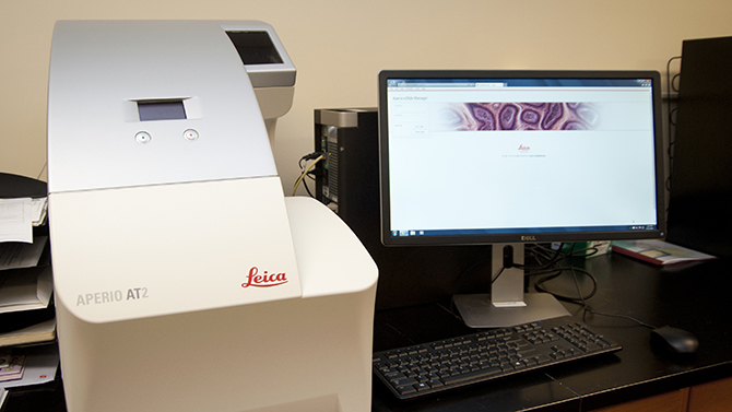
Digital pathology can go beyond the capabilities of a typical microscope where users are limited by objectives — the cylinders on a microscope that contain lenses for magnification of specimens — for a particular microscope. Digital pathology scanners available in the Comparative Pathology Laboratory include the Aperio AT2 brightfield scanner, which scans up to 40x objective, and the Versa, which is a fluorescence-based microscopy scanning system that scans up to 63x oil objective and up to six fluorescence channels.
A freely available image viewing and analysis system enables users to transition smoothly from a low-powered whole slide image to all magnifications between typical objectives so that users can orient themselves within the tissue and select the best magnification for the features they are hoping to view or analyze. The system also allows users to move freely around an image to find key areas and to run appropriate analytic algorithms.
“The system allows professors or researchers to annotate, that is to draw or mark right onto a specimen, so that they can point out key features to individuals either on the research or teaching level,” said Brannick.
With regard to the research applications, Brannick said the technology has already been used to collaborate internationally.
“Our very first test run allowed us to send an entire bank of slides for a research study to collaborators in Brazil, who were able to perform an analysis using the accompanying software and send data back to a professor here in completion of a thesis and toward a manuscript. We are really excited that the technology is already being put to the test and that it is already having a far-reaching effect,” said Brannick.
The technology also allows for seamless collaboration between researchers because slides are available virtually to the researchers and to anyone that the researcher permits to have access to those slides immediately after scanning.
The Aperio AT2 system also can accommodate scanning up to 200 slides at a time so whole research studies can be scanned within the course of an afternoon or evening. Images and associated data are stored virtually, making them accessible remotely anywhere with internet access. Important research or diagnostic case records can even be stored alongside virtual specimens so that important historical or clinical information is available to those accessing the specimens.
“You are not tethered to a scope. You are not tethered to a laboratory, and you are not tethered to the time restraints of a typical business work day. You can have access to your specimens when and where you desire,” said Brannick.
Brannick said digital pathology has broad uses and she can see it being utilized within and across many of UD’s seven colleges.
“We see this having broad applications for anyone working with plant and animal tissues, even human tissues, as well as for some researchers working at the cellular level – anyone that works with glass slides, really. We also see applications across biomedical research as well as basic life science research,” said Brannick.
She added another use is in archiving specimens, “as even older specimens can be scanned and stored indefinitely, virtually.”
Brannick, who said she is incredibly grateful for the funding from the Unidel Foundation that made it possible to bring this technology to campus, thinks that the biggest benefit to users is going to be the ability to access slides immediately upon scanning.
“You can conference in real time over an image so that you can work together to come to a diagnosis or a decision based on analysis. With the annotations on the research side, you could have one person measuring and scoring and leaving those annotations on that image and then another person could look at the same image without annotations, do their own scoring and compare. It is really versatile,” said Brannick.
About the Comparative Pathology Laboratory
The Comparative Pathology Laboratory, located at 127 Worrilow Hall, serves as a histology — tissue preparation — support laboratory as well as a pathology support laboratory for the whole University as needed.
Brannick, a certified veterinary anatomic pathologist, and Joanne Cooper, a certified histotechnician, can provide consultation to educators and researchers before, during, and after a project as well as offering specimen preparation, specimen scanning, and clinical or diagnostic interpretation of tissues.
Anyone interested in incorporating digital pathology into their research, teaching, or diagnostics should contact Brannick at brannick@udel.edu or visit the Comparative Pathology Laboratory website.
Contact Us
Have a UDaily story idea?
Contact us at ocm@udel.edu
Members of the press
Contact us at mediarelations@udel.edu or visit the Media Relations website