|
UNDERGRADUATE RESEARCH STUDENTS PRESENT THEIR WORK AT NATIONAL MEETINGS 2003-2004 |

|
|
|
UNDERGRADUATE RESEARCH STUDENTS PRESENT THEIR WORK AT NATIONAL MEETINGS 2003-2004 |
|
American Society
for Cell Biology
American
Chemical Society
Experimental
Biology Meetings
American Society for Biochemistry and Molecular Biology
American Society of Andrology
American Society
for Cell Biology Meetings
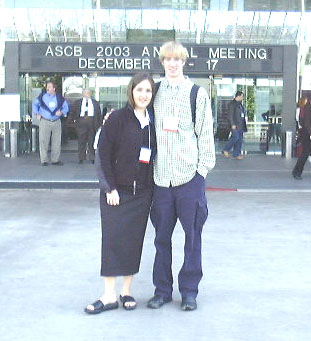
San Francisco, December 2003
|
Kathy Maddox and
Jim Parris presented their undergraduate research work at the Annual meetings
of the American Society for Cell Biology in San Francisco in December 2003. |
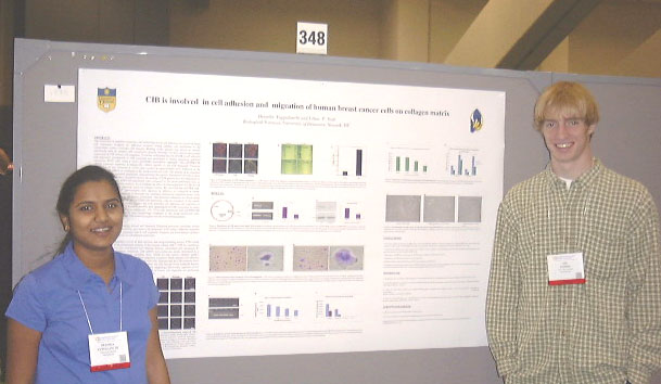
| A Novel Protein
that Interacts with Calcium and Integrin Binding Protein Induces Cellular
Adhesion and Spreading K. M. Maddox, M. U. Naik, K. Eckfeld, U. P. Naik; Biological Sciences, University of Delaware, Newark, DE 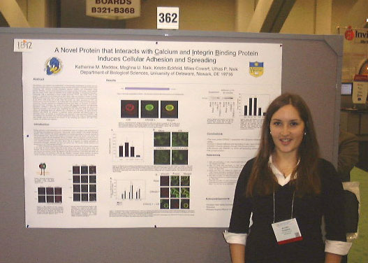
|
Cell
adhesion and migration are fundamental in understanding physiological
processes such as neo-vascularization and wound healing as well as several
pathological processes including tumor metastasis. Cell adhesion involves
interactions between integrins and extracellular matrix proteins. Recently,
we have shown that calcium and integrin binding (CIB) protein, which interacts
with the integrin αIIbβ3, plays an important role in platelet spreading
on immobilized fibrinogen. Additionally, CIB is expressed in human breast
carcinoma (T47D) cells where αIIbβ3 is not present. To determine the functional
role of CIB in these cells, we screened a human mammary epithelial cDNA
expression library in the yeast two-hybrid system and isolated a novel
polypeptide of 247 amino acids using CIB as bait. When this protein was
stably expressed in T47D and NIH3T3 cells a 2-3-fold increase in cell surface
area was observed using immunofluorescence microscopy. We also observed
an increased amount of stress fibers as visualized by phalloidin staining
of F-actin. Therefore, we named this protein CIB Regulated Adhesion and
Spreading Stimulator-1 (CRASS-1). When CIB was over expressed in CRASS-1
expressing cells we found about a 40% decrease in cell surface area, suggesting
that CIB inhibits the ability of CRASS-1 to induce cell spreading. Since
cell adhesion is a prerequisite for spreading, we asked whether CRASS-1 also
effects cell adhesion. In adhesion assays performed with NIH3T3 cells on
a fibronectin matrix, approximately a 2-fold increase in cell adhesion was
observed in cells expressing CRASS-1 and this adhesion was decreased by
40% when CIB was expressed in these cells. Thus, our studies suggest that
CRASS-1 may be involved in the induction of cell spreading and together
with CIB it may regulate cell adhesion and migration |
|
Developmental Expression Pattern of Junctional Adhesion Molecule-1 (JAM-1): Expression during Early Organogenesis J. J. Parris, P. B. Kelley, M. K. Duncan, U. P. Naik; Biological Sciences, University of Delaware, Newark, DE
|
Cell adhesion molecules of the Ig superfamily play an important role in embryonic development. We have recently shown that JAM-1, a member of this family, is involved in endothelial cell adhesion and migration leading to angiogenesis. This suggests that JAM-1 may also play a role in vasculogenesis; however, embryonic expression of JAM-1 is not well characterized. It has been reported using Northern analysis that JAM-1 transcript is not detected during the prenatal development of the mouse. In order to understand the role of JAM-1 in vascular development/function, we studied the expression of JAM-1 during early embryonic development by generating transgenic mice in which a lacZi gene was knocked in to the JAM-1 gene. JAM-1 gene activity was determined using histochemical staining for beta-galactosidase in mouse embryos ranging from 9.5 to 12.5 days post coitum (dpc). Contrary to previous findings, at 9.5 dpc there was a generalized expression of JAM-1 in the middle and posterior portions of the embryo, with high levels of expression in the otic pit, oral ectoderm, and Rathke’s pouch. By 10.5 dpc, the expression became localized to the mesonephros, hepatic/biliary primordial, midgut, lumen of the stomach, developing eye, olfactory epithelium, otocyst, and Rathke’s pouch. Expression was consistent in these structures as they developed; the heaviest expression occurred in Rathke’s pouch and the otocyst. At 12.5 dpc, additional staining was localized to the epithelial components of the metanephros and lungs as well as the choroid plexus and follicles of vibrissae. JAM-1 expression was also observed in the vasculature of whole mount embryos as well as in the atrial chamber of the heart and intersomitic vessels. Thus, JAM-1 may have a role during vasculogenesis and epithelial tissue development. |
|
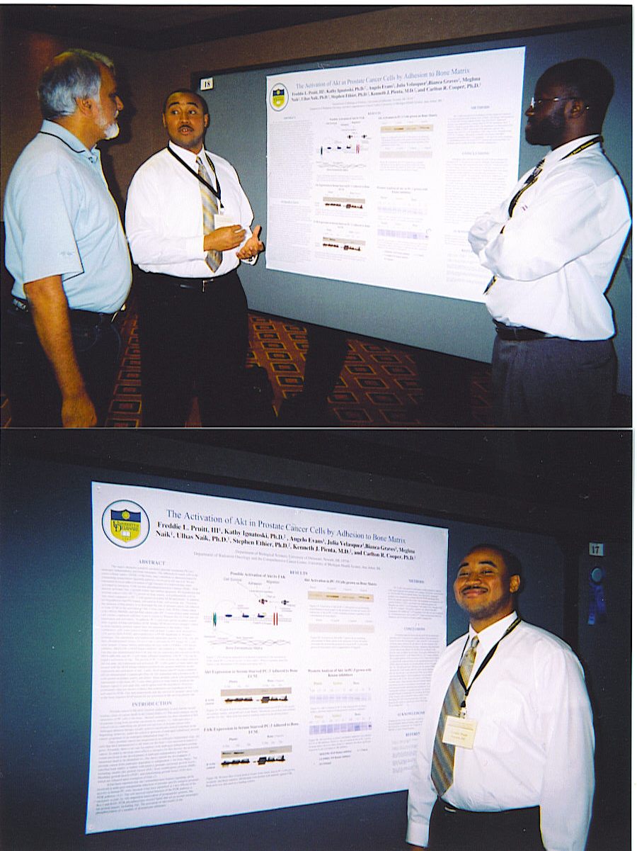
The Activation of Akt in Prostate
Cancer Cells by Adhesion to Bone Matrix. |
The
major obstacles posed by advanced prostate carcinoma (PC) are androgen
independence and bone metastasis. The adhesion of cancer cells to the extra-cellular
matrix (ECM) of the bone, may contribute to chemoresistance by stimulating
intracellular signaling pathways involved in cell survival. We are interested
in focal adhesion kinase (FAK) because it is believed that, when activated
by integrins, FAK recruits phosphatidylinositol 3-kinase (PI3K). PI3K
directly activates Akt, a protein kinase that inhibits apoptosis. We hypothesize
that prostate cancer cells (PC-3), grown on bone matrix, will preferentially
activate Akt when compared to PC-3 cells grown on kidney ECM and plastic.
In addition, we hypothesize that PI3 kinase, activated by FAK, will activate
Akt. Therefore, the purpose of this project is to determine the role
of prostate cancer cell adhesion to bone ECM in the activation of Akt.
Colon cancer cells (WiDr), breast cancer cells (MDA-MB468) and prostate
cancer cells (PC-3) were grown under standard cell culture conditions and
then lysed to screen by Western blot for FAK and Akt expression and activation.
In addition, PC-3 cells were grown on plates coated with 10μg/ml of bone
and kidney ECM. Kidney ECM served as a negative control to bone because
prostate cancer does not metastasize to the kidney. Upon confluency, cells
were lysed to isolate total proteins. Lysates were resolved in a 12% gel
by SDS-PAGE, and transferred to a PVDF membrane by Western technique. The
membrane was blotted with antibodies specific for FAK, Akt, and their phosphorylated
forms. To test if Akt is activated by PI3 kinase, PC-3 cells were treated
24 hours before performing the lysis with LY294002, a PI3 kinase inhibitor,
SB202190, a MAP kinase inhibitor. Our data has demonstrated that FAK and
Akt are expressed and activated in WiDr, MDA-MB-468, and PC-3 cells under
standard conditions, with PC-3 having the highest activation of Akt.
The growth of PC-3 cells on bone and kidney matrices did not alter Akt expression
and activation. PC-3 cells grown on bone matrix and treated with the MAP kinase
inhibitor produced the greatest inhibition on the expression and activation
of Akt. Since prostate cancer cells preferentially metastasize to the
bone, PC-3 cells when grown on bone matrix produced the highest signal of
activated Akt, which agrees with the hypothesis. MAP and PI3 kinase inhibitors
showed no significant effect on Akt activation in PC-3 cells grown on kidney
ECM and plastic. However, preliminary data has shown evidence that contradicts
our hypothesis of Akt activated by PI3K. Our data demonstrate that the survival
of prostate cancer cells in the bone requires MAP kinase for the activation
of the survival protein Akt. |
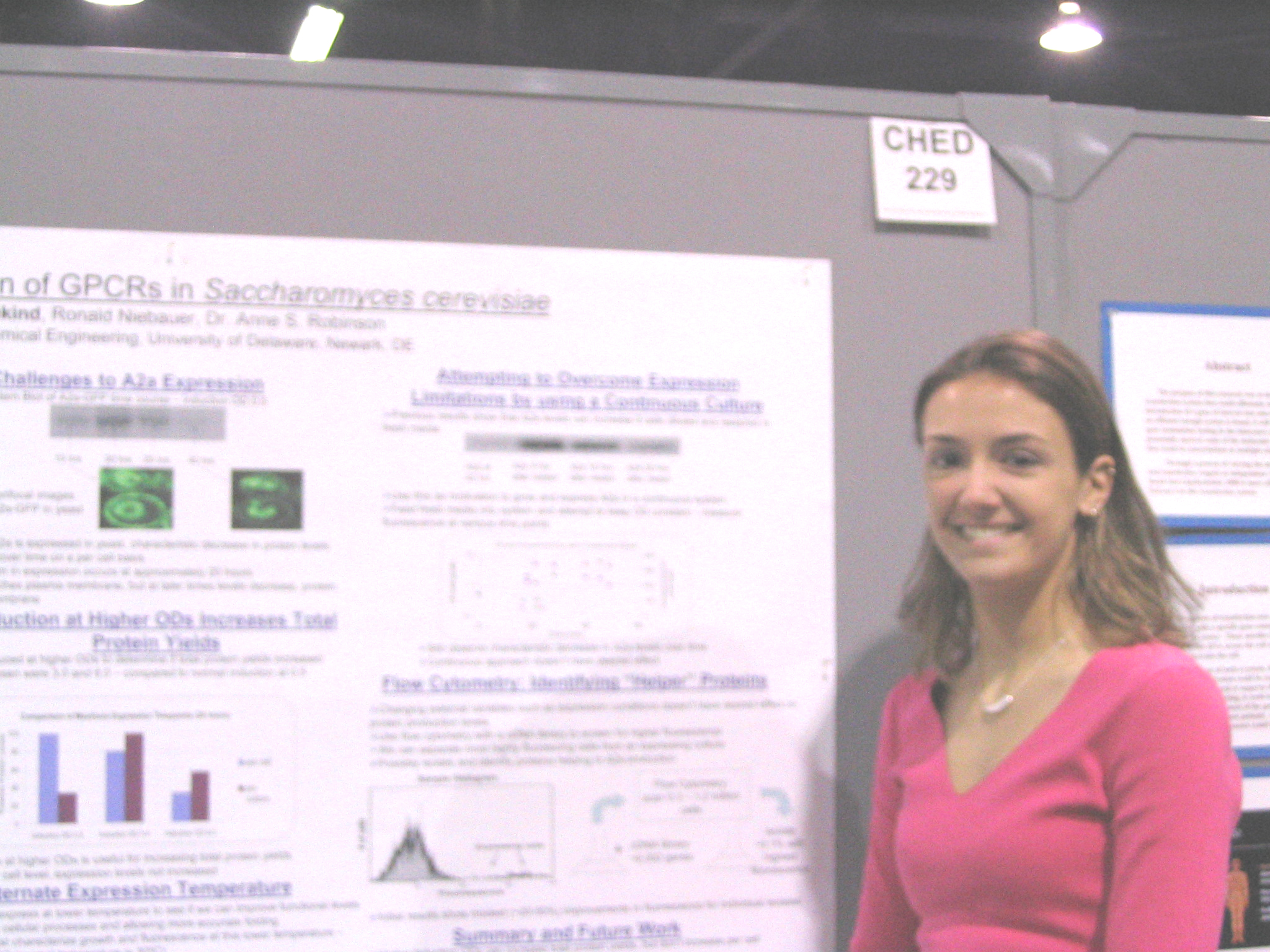
Optimizing Expression of GPCRs in Yeast |
Adenosine receptors are membrane proteins that play an important role in cell signaling and response through their interactions with adenosine. The adenosine receptors are a sub-family of the G protein-coupled receptor (GPCR) super-family. GPCRs have been implicated in many human diseases such as heart disease and asthma and are important drug targets. Knowledge about GPCR structure and function is limited by the difficulties associated with producing large amounts of functional protein for both biophysical and high resolution structural studies. Previous experiments in our laboratory have shown that when the adenosine receptor A2a is expressed in yeast, a systematic decrease in protein levels occurs over time. Thus, one of the research goals has been to gain insight into the causes of this decrease and to create an optimal system for A2a protein expression. For these studies, the A2a protein has been genetically fused to the green fluorescent protein (GFP), so that expression and trafficking may be monitored by fluorescence. We have found that several factors influence A2a expression, such as media conditions and expression temperature. Current studies with A2a expression include continuous culture growth, as well as optimizing induction and growth conditions to determine the effects on expression on a per cell as well as a per culture volume basis. |
Experimental
Biology Meetings
Washington, DC, April 2004
Expression Patterns of Orosomucoid During 3T3 L1 Adipocyte Differentiation Dept of Biological
Sciences, University of Delaware
|
The Ocular Expression and Regulation of Junctional Adhesion Molecule-1Dept. of Biological Sciences, University of Delaware, Newark, DE Junctional adhesion molecule-1 (JAM-1) is a member of the immunoglobin superfamily involved in the organization of tight junctions and the regulation of leukocyte transmigration. Recently, a cDNA microarray analysis of transgenic mice overexpressing PAX-6 in lens fiber cells revealed that JAM-1 mRNA expression was 2.5 fold elevated over normal. Thees data suggested that JAM-1 gene expression is regulated by PAX-6, a transcription factor essential for normal eye development. The overexpression of JAM-1 in the PAX-6 transgenic lenses of adult mice was confirmed by RT-PCR. A LacZ-Neo fusion genetrap was used to disrupt the JAM-1 gene in ES cells to create knockout mice and detect JAM-1 gene activity via B-galactosidase expression. In the lens and cornea, JAM-1 gene activity is detected in the epithelium, cells dependent on PAX-6 for normal morphogenesis. Levels decrease during lens fiber cell differentiation coincident with the downregulation of PAX-6 expression. Analysis of JAM-1 null mice revealed a down-regulation of B-galactosidase expression in the corneal epithelium relative to heterozygotes, suggesting JAM-1 may indirectly regulate its own expression. Further, histological analysis of aged JAM-1 null mice demonstrated that the corneal epithelia is thicker than normal and lacks a normal squamous layer; thus, JAM-1 is essential for normal corneal morphogenesis. Supported by the NIH. |
|
Expression of a Seventh
Hyaluronidase Gene, Hyal 5, in the Mouse 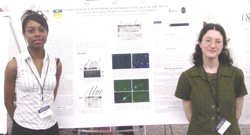
|
The
six mammalian hyaluronidase genes reside in two clusters located on chromosomes
3p and 7q in humans and 9 and 6A2 in the mouse. The three genes, HYALP1,
HYAL4, AND SPAM1 (PH-20) in the cluster on 7q/6A2 are all testis-expressed.
Of these, SPAM1, the Sperm Adhesion Molecule 1 has been best characterized
and has been shown to play multiple essential roles in mammalian fertilization.
It has recently been shown that a seventh hyaluronidase gene, Hyal5, which
is 60% homologous to the murine Spam1, is located approximately 100kb away
in the cluster and may be responsible for the fertility seen in Spam1 K/O
mice (Baba et al., JBC 2002). We have investigated the spatial and
temporal expression pattern of the rodent-specific Hyal5 in the mouse, as
a means of determining if the gene may have redundant or overlapping functions
with Spam1. Northern analysis shows a 1.5 kb transcript to be present
in the testis. Indirect immunofluorescence shows that the protein is
uniformly distributed on the heads of immature caput sperm while it is present
on the anterior and posterior head of caudal sperm, similar to the expression
of Spam1. The protein was also found to be present in the cytoplasmic
droplet of immature caput sperm. Further characterization of the protein
expression with indirect immunofluorescence and Western analysis is ongoing.
A developmental RT-PCR analysis was performed on mouse testes of 12, 18,
21, 22, 23, 25, and 27 post-natal day animals and the results indicate that
the transcript is post-meiotically expressed, similar to Spam1. Bioinformatical
data suggest that Hyal5 contains a cyclic AMP response element (CRE) in
its promoter region. Since several haploid-expressed testis-specific
genes have been found to have a CRE, this finding of a potential CRE element
is consistent with haploid expression of Hyal5. The findings suggest
that Hyal5 may be a redundant protein. It is important to note that
unlike Spam1, Hyal5 is not found in the human genome and therefore functions
performed by Hyal5 and Spam1 in the mouse are likely to be performed solely
by human SPAM1. Supported by NIH grant RO1 HD35158 to P.AM-D, Beckman Scholars, and Howard Hughes Medical Institute. |

|
| University
of Delaware HHMI-sponsored undergraduates and alumni met for dinner at the
Durgin Park restaurant in Boston June 12. From left front and clockwise around
the table: Isaac
Hubner (UD Biochemistry '01 now a graduate student in Chemistry
at Harvard) , Professor Hal White, Jake
LaPorte (UD Biochemistry '01 now a graduate student
in Chemistry at Harvard), Dan Oristian,
David
Nation, Mandy
Peters, Sarah
Redding, and Dan
Dries (UD Biochemistry '00 now a graduate
student in Pharmacology at the UC San Diego). |
|
Sarah Redding, Daniel Cline, Joel Schneider, and Colin Thorpe, Dept of Chemistry and Biochemistry, University of Delaware, Newark, DE |
The water-soluble phosphine tris(2-carboxyethyl)phosphine (TCEP) is now widely used as a stable disulfide reductant in a variety of applications in Biochemistry. Some studies report that TCEP is membrane pemeant, whereas others suggest that it cannot penetrate biological membranes. This poster examines the permeability of TCEP in comparison to other common comercial biochemical reductants, and reports the synthesis and characterization of new phosphine ester derivatives. Permeability was elevated with phospholipid vescicles containing entrapped DTNB (5,5'-dinitrobis(2-nitrobenzoic acd)) by following the increase in absorbance at 412 nm on reduction of the disulfide of DTNB. As expected, the highly polar tripeptide reductant glutathione shows no significant reduction of DTNB. We find that pure samples of TCEP are similarly impenetrant (as expected from its chemical structure). In contrast, a series of new ester analopgs was found to cross lipid bilayers very rapidly (considerably faster than dithiothreitol) suggesting that they may find use as modulators of redox-poise in cell biology. Supported in part by grants from HHMI and NIH GM26643. |
|
Cholesterol Efflux and Adipocyte Differentiation
Amanda Peters, John David, Jennifer
Risser, David C. Usher.
University of Delaware, Department of Biological Sciences, Newark, DE

|
Adipocytes
are known to be important regulators of fatty acid homeostasis.
The Serial Analysis of Gene Expression (SAGE) library of 3T3-L1 adipocytes,
constructed in our laboratory, has implicated cholesterol in having an
important role in this process. As determined by RT-PCR, the relative
expressions of several genes involved in cholesterol transport, Cav, SREBP-1c,
Abca1, and SR-B1, were found to be highly upregulated during 3T3-L1 differentiation.
To test the dependence of adipocytes differentiation on cholesterol in
forming their phenotypic lipid droplet, cholesterol efflux was induced
in cultured 3T3-L1 adipocytes on day 3 of differentiation, ensuring that
“early” genes, such as transcription factors, were allowed time for upregulation.
The addition of -cyclodextrin and HDL to the growth medium provided a
tool for cholesterol removal from 3T3-L1 cells without toxicity. Oil
red-O staining of mature 3T3-L1 cells after experimental treatment revealed
that cholesterol efflux during the late phase of differentiation greatly
hindered lipid droplet formation in adipocytes. This suggests that
cholesterol is necessary for lipid droplet development in adipocytes.
Expression patterns of “late” genes, including Cav, SREBP-1c, Abca1, SR-B1,
Adipsin, DGATI, CD36, PPAR, and Fabp4, were then compared to those under
conditions of cholesterol efflux. Funded by a grant from the Howard
Hughes Medical Institute.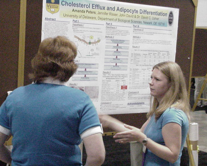
|
| Mandy Peters'
presenting her work in the Symposium on Cholesterol Homeostasis |
Mandy Peters
was awarded a First Prize in the ASBMB Undergraduate Poster Competition |
| HIP/RPL29 Function
in Early Mouse Embryos using Ribozyme Knockdown Daniel Oristian, Richard Focht, Catherine Kirn-Safran, Mary-Cindy Farach-Carson, Dept of Biological Sciences, University of Delaware, Newark, DE 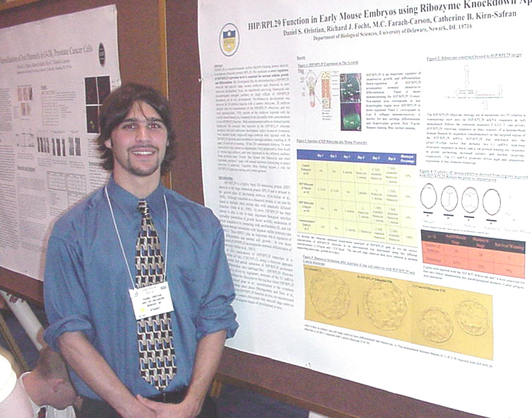
|
HIP/RPL29
is a heparin/heparin sulfate (Hp/HS) binding protein identical to peripheral
ribosomal protein L29. We postualte a strict regulation of HIP/RPL29 expression
level is essential for normal cellular growth and differentialtion.
We investigated this by microinjecting a HIP/RPL29 ribozyme inrto one-cell
stage mouse embryos and observed in vitro embryonic development. Next,
we transferred surviving blastocysts into pseudopregnant surrogate mothers
to study effects of HIP/RPL29 knockdown on in vivo development. Preblastocyst
development was observed for 10 embryos injected with a control ribozyme,
20 embryos injected with two concentrations of the HIP/RPL29 ribozyme, and
two were unmanipulated. 50% of the embryos injected with the control formed
blastocysts, compared to 10% for both concentrations of the HIP/RLP29 ribozyme.
Both unmanipulated embryos formed normal blastocysts. We conclude that
injection of the HIP/RLP29 ribozyme interferes with early embryonic develoment
before blastocyst formation. Single-cell stage embryos(220) were injected
with the HIP/RPL29 ribozyme and transferred to surrogate mothers, rsulting
in 18 pups; 16 survived for 4 days before re-implantation. 50 progressed
to form 4-cell to morula stage embryos, and were implanted into two different
mothers. None produced pups. 24 formed into blastocysts and when implanted,
produced 7 pups with normal skeletons. Genotyping to detect ribozyme is underway.
Together these findings support a role for HIP/RPL29 expression during early
embryogenesis. Supported by NIH (HD25235). |
|
Identification of ion
channels in C4-2B4 prostate cancer cells David
A. Nation, Kamil Akanbi, Mary C.
Farach-Carson. Dept. of Biological Sciences, University of Delaware,
Newark, DE 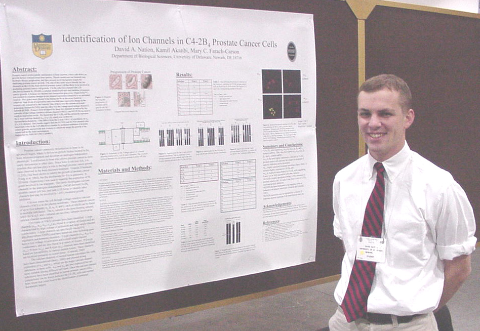
|
Prostate
cancer preferentially metastasizes to bone marrow, where cells thrive on
growth factors released from bone matrix. Plasma membrane ion channels
may facilitate disease progression, and thus present novel therapeutic targets
for inhibiting prostate cancer growth. The aim of this study was to
identify the ion channels in the C4-2B4 bone-derived prostate cancer cell
line likely to be involved in mediating prostate cancer cell growth.
C4-2B4 cells were treated with 1,25-dihydroxyvitamin-D3 (20 nM), a calcium
channel activator and inhibitor of prostate cancer growth. A human ion channel
and transporter gene array (SuperArray™ ) was probed to examine changes in
ion channel expression compared to an untreated control. Two genes
were chosen from among the 96 on the array based on relatively high levels
of expression and a two-fold plus expression change in the treated cells compared
to the control. One of these was the calcium-activated potassium channel
KCNN2, and the other was the voltage-gated sodium channel subunit SCN2B.
Primers were designed for these two channels as well as for the subunits
of the voltage-sensitive calcium channel (VSCC), and RT-PCR was used to confirm
expression levels. We found that the C4-2B4 cell line primarily expresses
the L-type calcium channel α1D [Cav1.3], which was verified by immunohistochemistry.
This differs from the L-type VSCC of osteoblasts, an α1C [Cav1.2] channel.
Our results suggest that the KCNN2 and SCN2B channels play important roles
in the 1,25-dihydroxyvitamin-D3 mediated inhibition of prostate cancer growth,
and provide new avenues to selectively target the growth of the cancer cell
in the bone environment. Supported by NIH/ NCI P01 CA098912. |
Travel and expenses for the above students
was provided primarily by the University of Delaware HHMI Undergraduate Science
Education Program with additional support from travel grants from the
American
Society for Biochemistry and Molecular Biology, the Beckman Scholars
Program, and the Women Scholars Program. The HHMI Program,
the Beckman
Scholars Program, Charles Peter White Fellowships, and the
Undergraduate Research Program supported
research by the students.