Abstracts from the Department of Biological Sciences
Undergraduate Summer Research Symposium August 9, 2006

Ordered alphabetically by student's last name

Ansbach Boyd Callahan Dennis Dumas Haag Hughes Kelleher
Awoyemi Brady, A Cheong Desai Engel Hadley Johnson,C Koemeter-Cox
Barninger Brady, M Chui Devulapalli Goldberger Herbert Johnson,L
Beringer Bruton Decker Dixon Grindel Higdon Kahn


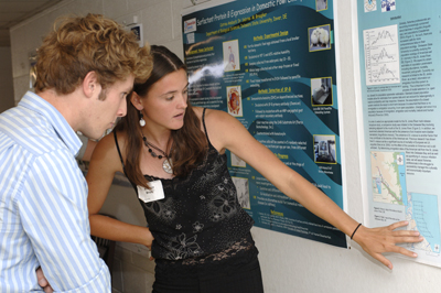
Surfactant Protein B Expression in Domestic Fowl Embryos

Catrina Ansbach
and Sabrina M. Brougher
Department of Biology, Delaware State University

Pulmonary surfactant is a mixture of phospholipids and proteins synthesized and secreted by alveolar type 2 (T2) cells.  While the functionality and composition of mammalian surfactant is well-documented, past research showing on avian surfactant protein is scarce.  The avian respiratory system differs from that of humans, yet they do share some similarities, as they both contain tubular structures (avian parabronchi and mammalian bronchioles) and share similar surfactant proteins (i.e. SP-A and SP-B).  Previous work shows embyonic expression of SP-A and SP-B, and, T2 cells were previously identified in hormone-cultured avian lung. However, further research is essential to understand the cellular mechanisms underlying T2 cell differentiation and SP-expression.  Based on the identification of these relevant physiological properties of avian lung tissue, or aim is that it will serve as a novel approach to biomedical research on human upper respiratory disease (i.e. asthma), with a specific interest in the identification and localization of SP-B. We hypothesize that an upregulation in the expression of SP-B will occur during embryonic development, in a T2-cell like histological pattern. Domestic fowl eggs were incubated, and a subset of 20 embryos were necropsied daily (from ed 10 through hatch). Harvested lung tissue was fixed in 4% PFA and paraffin-embedded. Tissue was sectioned and mounted on slides for immunohistochemistry. Immunohistochemistry will be performed for SP-B and expression quantified by counting the number of SP-B positive cells in 5 randomly selected fields of view per section (n=10 sections per age, from 10 different embryos). Supported by INBRE.


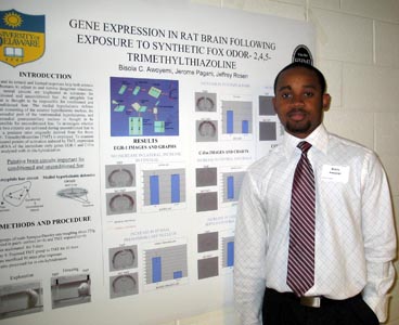
Gene Expression in Rat Brain Following Exposure to Synthetic Fox Odor – 2,4,5-Trimethylthiazoline

Bisola C. Awoyemi and  Jeffery B. Rosen
Department of Psychology

Two neural circuits are implicated as substrates for conditioned and unconditioned fear.  An amygdala fear circuit is thought to be responsible for conditioned and unconditioned fear.  The medial hypothalamic defense circuit consisting of the anterior hypothalamic nucleus, the dorsomedial part of the ventromedial hypothalamus, and dorsomedial premammillary nucleus is thought to be responsible for unconditioned fear. To investigate whether these two circuits are activated during unconditioned fear to a predator odor, the expression of immediate early genes EGR-1 and C-fos in brain was analyzed in rats after exposure to a synthetic predator odor originally derived from fox feces, 2,4,5-trimethylthiazoline (TMT). Two groups of male Sprague-Dawley rats, one control (n=4) and the other TMT-exposed (n=4), weighing about 225 g were housed in pairs - 12 hour light/ 12 hour dark cycle.  The rats were handled and acclimated to a test chamber for 15 min at about the same time for 3 days. On the 4th day, animals were acclimated for 5 min, exposed to TMT for 10 minutes and sacrificed 30 minutes after exposure. The brains were processed for in-situ hybridization. Increased EGR-1 mRNA expression was found in the TMT group when compared to the control group in many of the nuclei of the hypothalamic circuit.  Expression was not increased in the lateral amygdala. Expression of c-fos is still being processed. The results suggest that the hypothalamic circuit is the primary substrate for innate fear to predator odor. This work was supported by HHMI and NSF grant IBN-0129809. Acknowledgements: Jerome Pagani, Melanie Donley, McKenzie Herroon


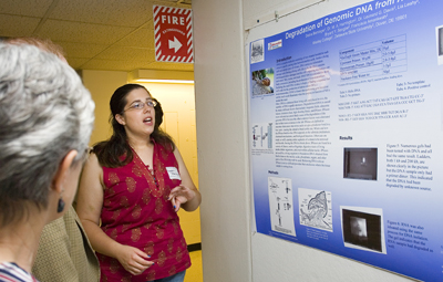
Degradation of Genomic DNA from Helix aspersa

Debra Barninger1,  M. A. Harrington2, Leonard G. Davis2, Lia Leahy2, Bryant Y. Sengbe2, Francisca Amankwah2
Wesley College1, Dept. Biological Sci., Delaware State University2, Dover, DE 19901

The goal of this research was to clone genes from the common garden snail Helix aspersa in the hopes of finding a gene responsible for neurotransmitter production using PCR. Genomic DNA is needed from the snails as a template. The primary purpose of DNA inside of a cell is information storage and DNA remains stable during the lifetime of the cell. DNA is repaired by the cell as necessary but generally remains unchanged throughout the life cycle of the cell. Once removed from a cell, DNA is highly prone to degradation from multiple sources including: magnetic fields, ultrasonic waves, extensive heat, high shearing forces, and DNases. The degradation greatly affected the ability to obtain and store genomic DNA from Helix aspersa.  A technical issue became the focus of the research to understand what could be causing the difficulties in storing genomic DNA. Possible sources of DNA degradation included evaluating laboratory conditions, preparation of all solutions used to obtain genomic DNA, and the isolation of tissue samples of snails from specific body locations. Once completed, proper steps were taken to ensure DNA stability. [Grant support NIH NCRR 2P2ORRO16472-04; NSF DBI-0320920 and IBN-0315551]


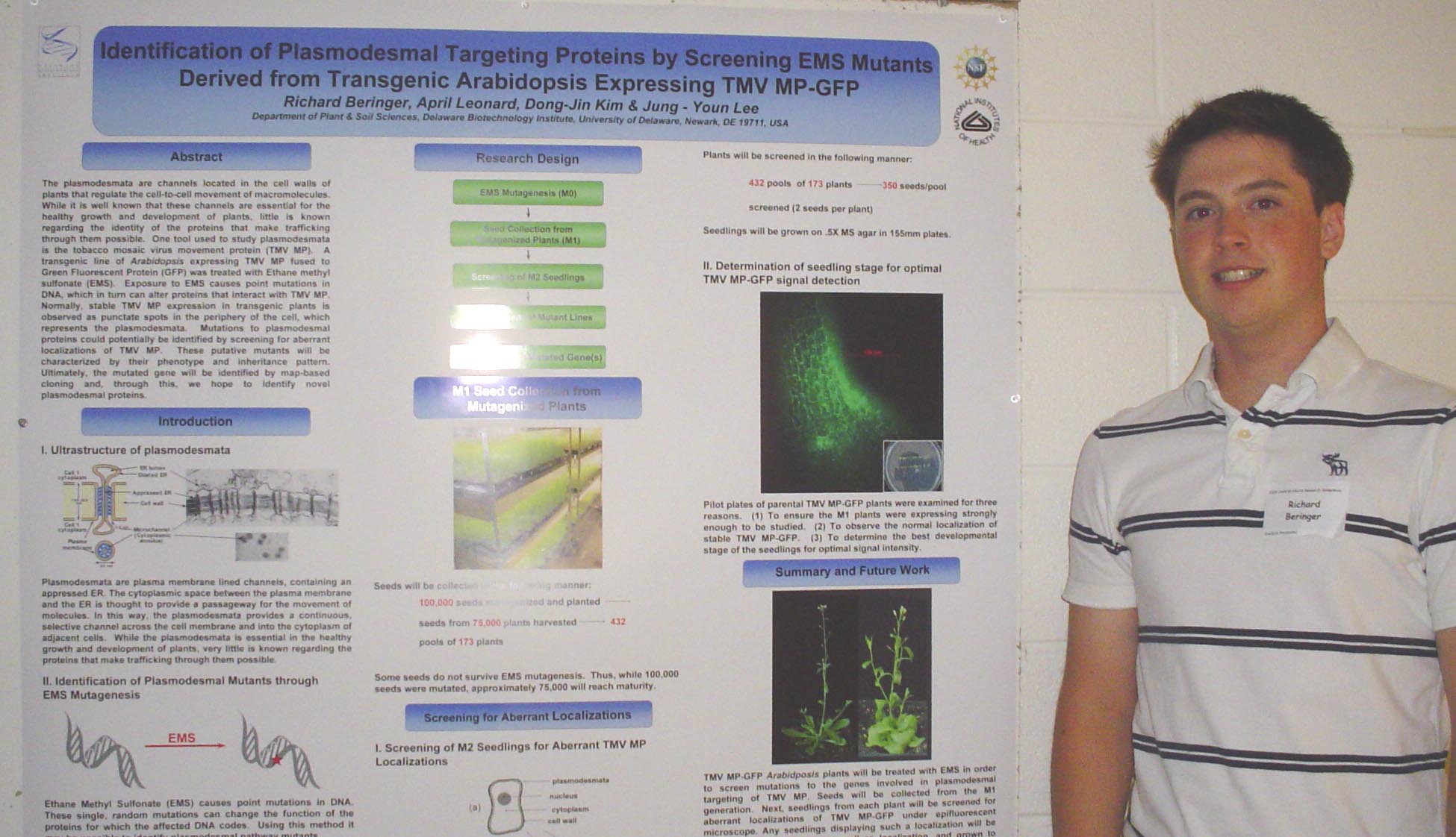
Identification of Plasmodesmal Targeting Proteins by Screening EMS Mutants Derived from Transgenic Arabidopsis Expressing TMVMP-GFP
Richard Beringer
, April Leonard, and Jung-Youn Lee
Department of Plant and Soil Science

The plasmodesmata are channels located in the cell walls of plants that regulate the cell-to-cell movement of macromolecules.  While it is well known that these channels are essential for the healthy growth and development of plants, little is known regarding the identity of the proteins that make trafficking through them possible.  One tool used to study plasmodesmata is the tobacco mosaic virus movement protein (TMVMP).  A transgenic line of Arabidopsis expressing TMVMP fused to Green Fluorescent Protein (GFP) was treated with Ethane methyl sulfonate (EMS).  Exposure to EMS causes point mutations in DNA, which in turn can alter proteins that interact with TMVMP.  Normally, stable TMVMP expression in transgenic plants is observed as punctate spots in the periphery of the cell, which represents the plasmodesmata.  Mutations to plasmodesmal proteins could potentially be identified by screening for aberrant localizations of TMVMP.  These putative mutants will be characterized by their phenotype and inheritance pattern.  Ultimately, the mutated gene will be identified by map-based cloning and, through this, we hope to identify novel plasmodesmal proteins.  Funding provided by the University of Delaware Undergraduate Research Fellows Program.


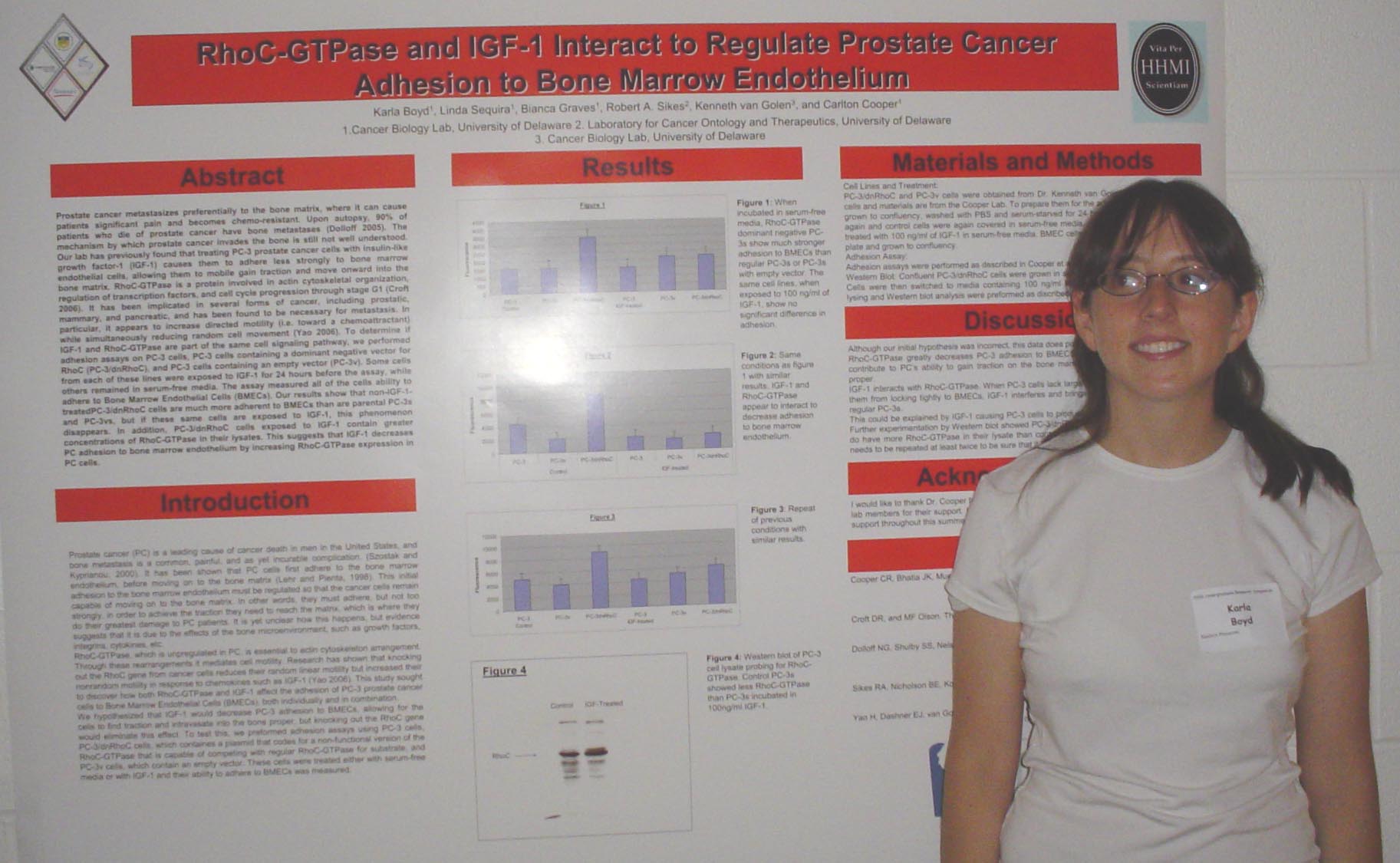
RhoC-GTPase and IGF-1 Interact to Regulate Prostate Cancer Adhesion to Bone Marrow Endothelium
Karla Boyd
, Linda Sequira, Bianca Graves, Kenneth van Golen, and Carlton Cooper
Department of Biological Sciences


Prostate cancer metastasizes preferentially to the bone matrix, where it can cause patients significant pain and becomes chemoresistant. Upon autopsy, 90% of patients who die of prostate cancer have bone metastases. The mechanism by which prostate cancer invades the bone is still not well understood. Our lab has previously found that treating PC-3 prostate cancer cells with Insulin-like Growth Factor-1 (IGF-1) causes them to adhere less strongly to bone marrow endothelial cells, allowing them to gain traction and move onward into the bone matrix. RhoC-GTPase is a protein involved in actin cytoskeletal organization, regulation of transcription factors, and cell cycle progression through stage G1. It has been implicated in several forms of cancer, including prostatic, mammary, and pancreatic, and has been found to be necessary for metastasis. In particular, it appears to increase directed motility (ie toward a chemoattractant) while simultaneously reducing random cell movement. To determine if IGF-1 and RhoC-GTPase are part of the same cell signaling pathway, we performed adhesion assays on PC-3 cells, PC-3 cells containing a knockout vector for RhoC (PC-3/dnRhoC), and PC-3 cells containing an empty vector (PC-3v). Some cells from each of these lines were exposed to IGF-1 for 24 hours before the assay, while others remained in serum-free media. The assay measured all of the cells ability to adhere to Bone Marrow Endothelial Cells (BMECs). Our results show that non-IGF-1-treatedPC-3/dnRhoC cells are much more adherent to BMECs than are PC-3s or PC-3vs, but if these same cells are exposed to IGF-1, this phenomenon disappears. This suggests that IGF-1 may upregulate the cells own manufacturing of RhoC-GTPase. Supported by the HHMI Undergraduate Science Education Program.


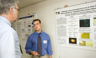
Modeling Tumor Development in an In Vivo Chick Embryo Model for Targeting with Carbon Nanotubes

Adam Brady1, Deni S. Galileo1, Balaji Panchapakesan2
Departments of 1Biological Sciences and 2Electrical Engineering

Carbon nanotubes (CNTs) are synthetic allotropes of sp2 hybridized carbon atoms in a cylindrical configuration.  CNTs possess many novel properties, including the ability to explode when clusters of CNTs are irradiated with light.  This property shows promise as a future therapeutic in the fight against cancer.  However, CNT treatments are far from reality and much testing is needed to determine how they interact within an organism.  We are developing a series of models of tumor formation using tumor cell lines and early chicken embryos.  Our first model consists of LacZ positive rat glioma cells injected directly into the vasculature of the chick embryos.  The tumors were visualized using X-Gal which stains LacZ positive cells blue.  The next model involves a shell-less embryo culture with plugs consisting of Matrigel, basic fibroblast growth factor (bFGF), and glioma cells resting directly on the chorioallantoic membrane of the embryo.  In addition to the tumor models, we non-covalently bound CNTs to biotinylated marker antibodies and injected them into the vasculature of chick embryos.  This served as a preliminary toxicity test as well as a potential way to visualize the distribution of CNTs in vivo.  In the future, shell-less embryos will be used to culture surgical explants of human brain and breast cancers, followed by their injection with CNTs.  These models will help to simulate brain tumor formation and will serve as a testing ground for in vivo targeting of CNTs to cancer cells.  This project was supported by NIH-NCRR-INBRE and HHMI.


An Assessment of American Eel (Anguilla rostrata) Dispersal and Prevalence of Parasitism
by Anguillicola carassus in the Waters Surrounding the St. Jones River, Delaware

Marissa G. Brady, Colette M. Cairns, and Dewayne Fox
Delaware State University

The American eel (Anguilla rostrata) ranks as one of the most important commercial fishes in Delaware which when coupled with reported population declines is problematic. This two-part study was conducted to determine if American eel emigrated from the St. Jones River, Delaware into nearby waters of the Delaware Bay as well as the prevalence of an invasive parasite, Anguillicola crassus.  During June -July ,2006 we placed 36 commercial eel pots in Delaware Bay waters adjacent to the mouth of the St. Jones River assess emigration from the river.  Using GIS mapping software, fixed sampling locations were set in the Delaware Bay. Commercial eel pots were allowed to fish overnight and captured American eel were scanned for the presence of passive integrated transponder (PIT) tags. A total of 79 American eel were collected, of which three (3.8%) were previously tagged in the St. Jones River.  This low number of recaptures in Delaware Bay indicates that there is limited dispersal of American eel between the St. Jones River and Delaware Bay. We also examined American eel for the presence of A. crassus, an invasive nematode that infects the swim bladder. Our results indicate a large incidence of infection rates (38%) for this non-native parasite. This infection rate is higher compared to previously published work (e.g. Chesapeake Bay (10-29%), Hudson River (0-12%)) and is problematic for the long term sustainability of the population. Our results are important as fisheries professionals struggle to successfully manage this culturally and economically important resource. Funded by EPSCoR.


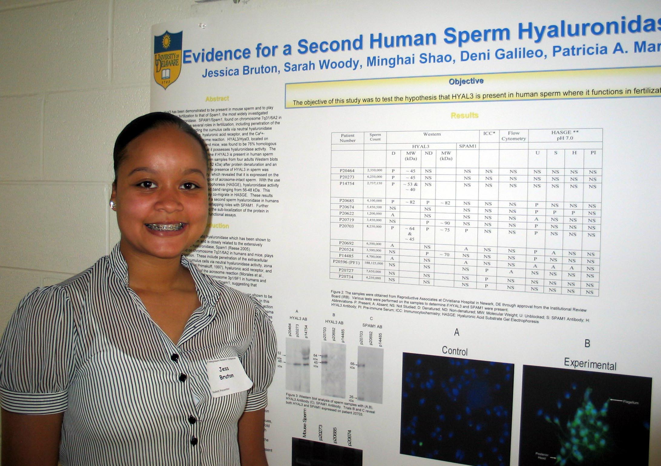
Evidence for a Second Human Sperm Hyaluronidase: HYAL3

Jessica Bruton, Sarah Woody, Minghai Shao, Deni Galileo, Patricia A. Martin-DeLeon
Department of Biological Sciences

Hyal3 has been demonstrated to be present in mouse sperm and to play similar roles in fertilization to that of Spam1, the most widely investigated reproductive hyaluronidase.  SPAM1/Spam1, found on chromosome 7q31/6A2 in humans and mice, plays several roles in fertilization, including penetration of the extracellular matrix surrounding the cumulus cells via neutral hyaluronidase activity, zona pellucida binding, hyaluronic acid receptor, and the Ca²+-associated induction of the acrosome reaction.  HYAL3/Hyal3, located on chromosome 3p1/9F1 in humans and mice, was found to be 76% homologous with SPAM1/Spam1; suggesting that it possesses hyaluronidase activity.  The objective of this study was to determine if HYAL3 is present in human sperm where it is functionally active.  In sperm samples from four adults Western blots revealed 2 isoforms (~ 42 kDa and ~ 48 kDa) after protein denaturation and an ~ 82 kDa HYAL3 (non-denatured).  The presence of HYAL3 in sperm was confirmed with immunocytochemistry, which revealed that it is expressed on the posterior head and the equatorial region of acrosome-intact sperm.  With the use of hyaluronic acid substrate gel electrophoresis (HASGE), hyaluronidase activity was identified at neutral pH as a broad band ranging from 56-48 kDa.  This suggests that SPAM1 and HYAL3 may co-migrate in HASGE.  These results show for the first time the presence of a second sperm hyaluronidase in humans and suggest that HYAL3 may play overlapping roles with SPAM1.  Further studies will be focused on determining the sub-localization of the protein in acrosome-reacted sperm and its role in functional assays. Funded by IGERT.


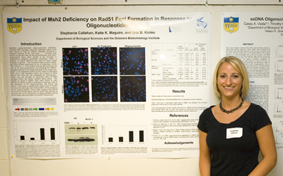
Impact of Msh2 Deficiency on Rad51 Foci Formation in Response to Oligonucleotides

Stephanie Callahan, Katie Maguire,
and Eric Kmiec
Department of Biological Sciences and the Delaware Biotechnology Institute

Modified single-stranded oligonucleotides (MSSOs) activate DNA damage response pathways within cells and induce a host of cellular changes including a transient stalling of the cell cycle.  Proteins with known functional roles as anti-recombinases and tumor suppressors have been shown to be involved in suppressing targeted nucleotide exchange (TNE), a process in which MSSOs are used to direct base changes in genes.  The mismatch repair protein, Msh2, has been shown previously to be a suppressor of the TNE reaction.  This protein may be acting through its role as an anti-recombinase by inhibiting the Rad51-mediated pairing of the oligonucleotide to the target site. Rad51 is an essential protein for the repair of damaged DNA and it will relocalize within the nucleus to form distinct foci that can be visualized by microscopy.  These foci are thought to represent sites of DNA damage where the repair reaction takes place.  We wanted to know if Rad51 was more active in cells lacking the Msh2 protein compared to wild-type cells in the presence of an oligonucleotide.  To address this question we electroporated Msh2 deficient and wild-type cells in the presence and absence of the oligonucleotide and determined the level of Rad51 foci formation by confocal microscopy.  We found that Msh2-/- cells have a significantly higher level of Rad51 foci then the wild-type cells independent of the presence of the oligonucleotide.  This data suggests that cells missing the Msh2 protein are more recombinogenic and as such may provide an environment more conducive to oligonucleotide pairing at the target site and higher correction frequencies. Funded in part by INBRE.


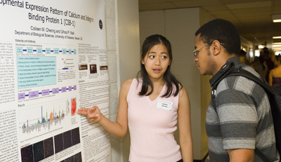
Developmental Expression Pattern of Calcium and Integrin-Binding Protein-1 (CIB-1)
Colleen M. Cheong and Ulhas P. Naik
Department of Biological Sciences

Calcium and integrin binding protein-1 (CIB-1) is known to have a key function in platelet activation through its interaction with integrin αIIbβ3.  Through Northern blots, it has also been found that CIB-1 mRNA is present in a wide number of adult tissues, including heart and kidney.  However, the localization of CIB-1 in these tissues and the role of CIB-1 during development have yet to be studied.  In order to study the expression pattern of CIB-1, a series of immunofluorescence experiments were performed.  First, the immunohistochemical staining protocol for the staining of CIB-1 was standardized.  Next, sections of heart and kidney tissue were stained and viewed using fluorescence microscopy.  From these experiments, it was found that CIB-1 protein is expressed in adult mouse heart and kidney tissues.  From the expression patterns, it appears that CIB-1 is expressed in the blood vessels of the heart and in the periphery of the glomeruli of the kidney.  Further experimentation is necessary to conclusively determine this expression pattern.  Additionally, mouse embryos were collected from 9.5-16.5 days post coitum (d.p.c.) through timed-pregnancies. Some of these embryos have been sectioned and stained to examine the contribution of CIB-1 in developing organs.  Furthermore, a non-radioactive in situ hybridization protocol is being standardized in order to study the expression pattern of CIB-1 mRNA.  From these experiments, we will gain better understanding of the overall expression pattern of CIB-1 in various tissues, which may give some indication of the function of CIB-1.  This research was supported by the Howard Hughes Medical Institute.


Expression of Heparan Sulfate Remodeling Enzymes,

Heparan Sulfate 6-O-Endosulfatases, in the Pregnant Mouse Uterus
Cecilia Chui 1, Sonia D'Souza 2, Mary C. Farach-Carson 2 and Daniel D. Carson 2
1 Imperial College London, London, United Kingdom , 2 Department of Biological Sciences, University of Delaware, Newark, Delaware


Heparan sulfate (HS) 6-O-endosulfatases (Sulfs) represent a recently discovered family of enzymes that are secreted via the Golgi and bind to the cell surface or are released into the extracellular matrix (ECM). Sulfs remodel HS proteoglycans (HSPGs), growth factor binding components of the ECM. These enzymes remove the 6-O sulfate group from trisulfated disaccharides of HS chains at neutral pH. By editing the sulfation status of these highly sulfated motifs, the specificity and affinity of interactions between HS and molecules such as growth factors and morphogens are modulated. Two Sulf family members, Sulf-1 and Sulf-2, have been described in diverse species including quail, rat, mouse and human. Though Sulf-2 is very similar in structural organization to Sulf-1, amino acid sequence identity is divergent (65%) between mouse and human. Both enzymes are expressed in multiple tissues, but with different relative levels of expression among tissues. Sulf expression has never been examined in the context of the uterus, even though HSPGs play critical roles in growth processes occurring in implantation and pregnancy. The objective of this project was to study the expression of both Sulf-1 and Sulf-2 in pregnant mouse uterus through RT-PCR and Q-PCR analysis. Primers were designed to amplify DNA coding for the enzymes of interest.  Initial experiments were conducted using total mouse embryo RNA to optimize the conditions for PCR using Sulf-1 and Sulf-2 primers. Subsequently cDNA reversely transcribed from RNA extracted from mouse uterus during early stages of pregnancy was used as template in PCR to examine the expression of mRNA encoding these enzymes. Results of the research will give an insight on the potential role(s) of these enzymes play in modulating HS binding growth factory delivery in early pregnancy. (Supported by University of Delaware/Imperial College Undergraduate Research Exchange Program and NIH grant HD25235 to D.D.C). 


βB2-crystallin regulation of the lens cytoskeleton

Corinne Decker, Kevin Duprey, Yan Wang, and Melinda Duncan
Department of Biological Sciences

The vertebrate lens is an avascular, transparent tissue composed of epithelial cells and fiber cells.  Both cell types contain crystallins, a class of proteins necessary to maintain the high refractive index of the ocular lens.  βB2-crystallin is the most prevalent β-crystallin in the adult lens; damaged βB2-crystallin aggregates contribute to natural cataract formation.  Lens transparency is also dependent upon cytoskeletal elements such as actin, tubulin, and the beaded filament, composed of CP49 and filensin.  This investigation seeks to assess the effect of βB2-crystallin expression on the lens cytoskeleton.  The Philly mouse, a strain characterized by a mutated βB2-crystallin gene and consequent inherited cataract will be used to complete this analysis. Expression of filamentous actin appears normal in Philly mutant and heterozygote newborn fiber cells, which are also characterized by positive immunohistochemical staining for CP49 and filensin.  One month after birth, mutant fiber cells are void of f-actin, while expression of CP49 and filensin is upregulated.  Both wild type and mutant newborns stained insignificant levels of α-smooth muscle actin.  Epithelial cells of one month-old Philly mutants are characterized by an upregulation of α-smooth muscle actin when compared to age-matched wild-type mice, a sign that epithelial-mesenchymal transition, a wound-healing response, is taking place in these cells.  These results indicate that the cytoskeleton appears normal at birth, and changes in the cytoskeleton occur as a result of the postnatal onset of βB2-crystallin expression.  Upcoming experiments include wholemount immunostaining for tubulin, as well as analysis of β-B2 crystallin expression in newborn to adult mice by PCR techniques.  Funded by Howard Hughes Medical Institute.


Developing a Student Educational Trail for Delaware State University’s Wetland

Yyone Dennis, Leonard Davis, Richard Driskill, Susan Yost, and Gary Kreamer
Biology Department, Delaware State University, 1200 N. DuPont Highway, Dover, Delaware 19904.

The wetland studied is located on the DSU campus with its water connected to Fork Branch, a stream leading into Silver Lake. A wetland is a low area where the land is saturated by water, but contrary to popular belief, wetlands are not always visibly wet; in such cases plants and soil types are the defining wetland indicators. In order to develop an educational trail, our first step was to document the historical impact of humans and then to remove all the trash.  Water and soil tests were performed to establish the ‘health’ of the wetland, as well as the dynamics of the soil-water interactions.  Data was collected to identify the water-flow and capacity before and after a rainfall.  With that information, the layout of the educational trail was planned.  This trail will include several stations, offering observational as well as interactive learning experiences focusing on wetland characteristics and values, human impacts, and aspects of the wetland flora and fauna.  These stations will include pictorial information on plants and animals that inhabit the area.  This project has successfully collected the baseline data and established a plan to achieve the goal of establishing a nature trail that would serve to both educate and inspire its trail-takers.  The next phase will be to construct and implement the structural components.  We have secured the support of Boy Scouts and DSU engineering staff to assist in constructing a stairway into the wetland, signposts and a trail system that loops around the wetland area. Supported by NSF EPSCoR Grant No. EPS-0447610.


CD44 and Posterior Capsular Opacification

Vivek D. Desai
and Melinda K. Duncan
Department of Biological Sciences


Posterior capsular opacification (PCO) is an undesirable wound healing response in which the residual lens cells remaining in the eye after cataract surgery proliferate, migrate into the visual field, and synthesize extracellular matrix molecular similar to those found in scar tissue, damaging the patient’s vision. PCO arises from epithelial mesenchymal transition (EMT) of lens epithelial cells. In other systems, CD44, a receptor for hyaluronan, has been identified to mediate changes in cellular proliferation, migration and cell identity leading to EMT. We hypothesize that the exposure of lens cells to hyaluronan based viscoelastics stimulates PCO following extracapsular lens extraction and that the hyaluronan receptor CD44 is involved in EMT of lens cells. In the adult mouse lens, CD44 is expressed in the lens fiber cells and not expressed in the lens epithelial cells, whereas in embryonic mouse lens, CD44 is not expressed in both the lens fiber cells and the lens epithelial cells. RT-PCR demonstrated that the “canonical” version of CD44 is the major CD44 splice variant in the lens. In adult mice, CD44 is not expressed in the lens epithelial cells immediately after the cataract surgery, however, its expression highly up-regulates in the lens epithelial cells 1-day following the surgery. Because of the dynamics of CD44 during the development of mouse lens and following the cataract surgery in adult mouse lens, out immediate future work is to study the distribution of CD44 ligands like hyaluronan and osteopontin. Supported by a Beckman Fellowship.


Effect of Neurotoxic Lesions of the Basolateral Complex of the Amygdala

on Contextual Fear Conditioning to a Predator Odor
Chris Devulapalli and Jeffrey B. Rosen
Department of Psychology 

Significant evidence supports the notion that the basolateral complex of the amygdala plays a role in unconditioned fear and contextual fear conditioning induced by foot shock. However, recent evidence argues that neurotoxic lesions to the basolateral complex significantly reduces conditioning to painful foot shock but does not affect the unconditioned fear response to a non-painful predator odor Trimethylthiazoline (TMT). To investigate whether the basolateral complex plays a role in contextual fear conditioning to TMT, neurotoxic lesions of the basolateral complex were made in rats. Contextual fear conditioning with TMT was then conducted on these lesioned rats and a non-lesioned control group. This procedure involved placing the rats into a chamber with a hide box for acclimation, and then on the next day exposing them to TMT in the chamber for 15 minutes. Finally, a memory test was performed 24 hours after exposure to the TMT by placing the rats back into the chamber without any TMT. Fear behaviors such as freezing (immobility) and time spent in the hide box were measured. It is anticipated that the lesioned animals will show significantly less conditioning to the chamber than the non-lesioned animals, as measured by less freezing and time spent in hide box during the memory test. These results would suggest that the basolateral complex of the amygdala is involved in fear conditioning to several different kinds of unconditioned fear stimuli such as foot shock and predator odor, but is not important for unconditioned fear responses to predator odor. This work was supported by Howard Hughes Medical Institute & National Science Foundation Grant IBN- 0129809


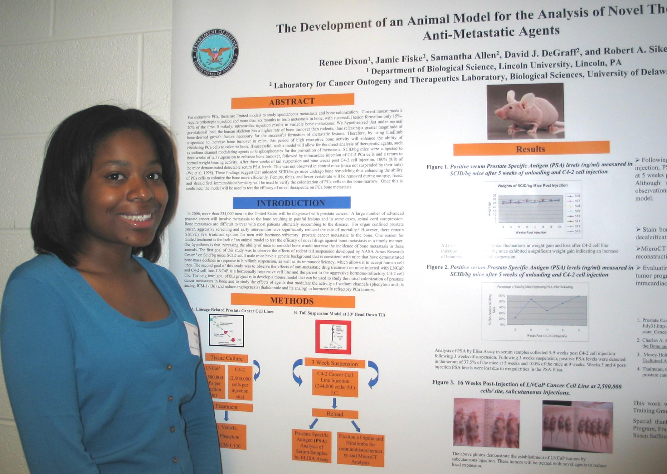
The Development of an Animal Model for the Analysis of Novel Therapeutic Anti-Metastatic Agents

Renee Dixon, 1 Jamie Fiske, 2 Samantha Allen, 2 David J. DeGraff, 2 and Robert A. Sikes 2
1 Department of Biological Science, Lincoln University, Lincoln, PA
2
Cancer Ontogeny and Therapeutics Laboratory in the Department of Biological Sciences, University of Delaware, Newark, DE

There are limited models to study the spontaneous metastasis and bone colonization of prostate cancer (PCa).  Current mouse models require orthotopic injection and more than six months to form metastasis in bone, with successful lesion formation only 15%-20% of the time. Similarly, intracardiac injection results in variable bone metastases. We hypothesized that under normal gravitational load, the human skeleton has a higher rate of bone turnover than rodents, thus releasing a greater magnitude of bone-derived growth factors necessary for the successful formation of metastatic lesions. Therefore, by using hindlimb suspension to increase bone turnover in mice, this period of high resorptive bone activity will enhance the ability of circulating PCa cells to colonize bone. If successful, this model will allow for the direct analysis of therapeutic agents, such as sodium channel modulating agents or bisphosphonates on cancer metastasis. SCID/bg mice were subjected to three weeks of tail suspension, followed by intracardiac injection of C4-2 PCa cells and a return to normal weight bearing activity. Nine weeks post injection, 100% (8/8) of the mice demonstrated detectable serum PSA levels. These findings suggest that unloaded SCID/beige mice undergo bone remodeling thus enhancing the ability of PCa cells to colonize the bone. Funding by the Department of Defense HBCU/MI Undergraduate Research Training Grant, PC05136.


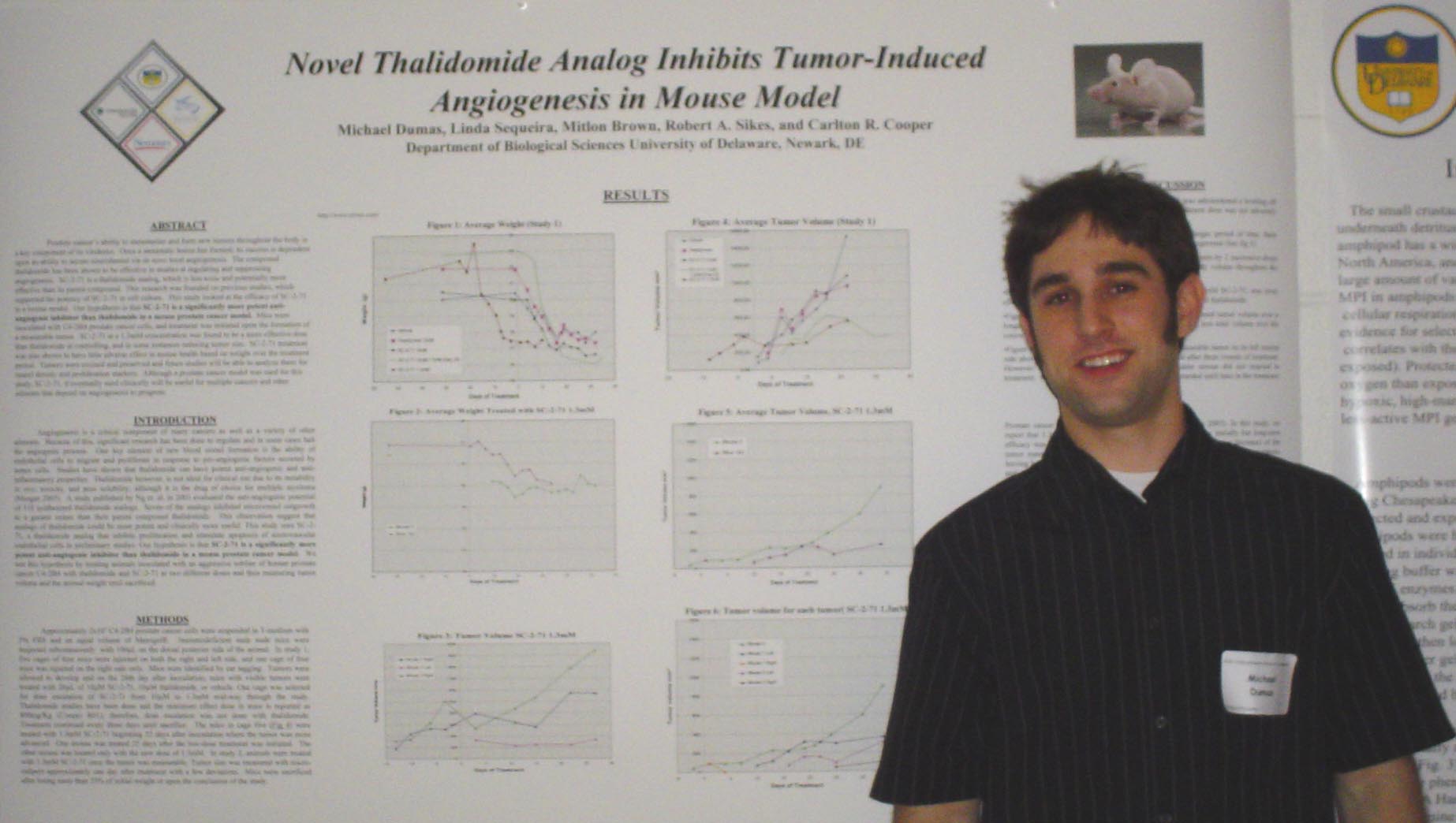
Novel Thalidomide Analog Inhibits Tumor-Induced Angiogenesis in Mouse Model

Michael Dumas, Linda Sequeira, Robert A. Sikes, and Carlton Cooper
Department of Biological Sciences

Prostate cancer’s ability to metastasize and form new tumors throughout the body is a key component of its virulence.  Once a metastatic lesion has formed; its success is dependent upon its ability to secure nourishment via de novo local angiogenesis.  The compound thalidomide has been shown to be effective in studies at regulating and suppressing angiogenesis.  SC-2-71 is a thalidomide analog, which is less toxic and potentially more effective than its parent compound.  This research was founded on previous studies, which supported the potency of SC-2-71 in cell culture.  This study looked at the efficacy of SC-2-71 in a mouse model.  Our hypothesis is that SC-2-71 is a significantly more potent anti-angiogenic inhibitor than thalidomide in a mouse prostate cancer model.  Mice were inoculated with C4-2B4 prostate cancer cells, and treatment was initiated upon the formation of a measurable tumor.  SC-2-71 at a 1.3mM concentration was found to be a more effective dose than thalidomide at controlling, and in some instances reducing tumor size.  SC-2-71 treatment was also shown to have little adverse effect in mouse health based on weight over the treatment period.  Tumors were excised and preserved and future studies will be able to analyze them for tunnel density and proliferation markers.  Although a prostate cancer model was used for this study, SC-2-71, if eventually used clinically will be useful for multiple cancers and other ailments that depend on angiogenesis to progress.


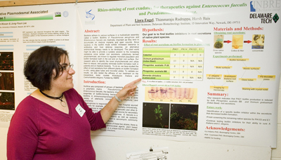
Rhizo-mining of Root Exudates for Therapeutics Against Enterococcus faecalis and Pseudomonas aeruginosa
Liora Engel, Thimmaraju Rudrapa, Harsh Bais
Department of Plant and Soil Science

Bacteria adhere to various surfaces in a multicellular assembly called a biofilm. Biofilms of Pseudomonas aeruginosa and Enterococcus faecalis are medically important as they tend to accumulate on medical implants and open wounds. Since bacteria in the biofilm mode exhibit increased tolerance to antibiotics and host defense response, an alternative antimicrobial therapy needs to be discovered. The plant world might be able to provide a suitable solution to the increasing resistance since certain root secretions, collectively termed phytochemicals, are known to regulate microbial population and biofilm formation both in the soil and on their root surface. Our research aims to identify the novel phytochemicals and utilize them to reduce, and possibly eliminate biofilm formation both in patients and on medical implants.  To do so, we have studied the effect of root secretions from different plants on biofilm formation in-vitro, using glass tubes and microtiter plates. To validate our results, we also tested the efficacy of our treatment on the inexpensive biotic models Arabidopsis and C. elegans. Funded by INBRE.


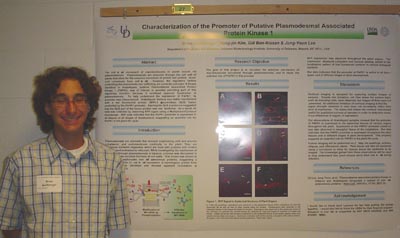
Characterization of the Promoter of Putative Plasmodesmal-Associated Protein Kinase 1

Brian Goldberger
and Jung-Youn Lee
Department of Plant and Soil Science

The cell-to-cell movement of macromolecules in plants occurs via plasmodesmata.  Plasmodesmata are channels through the cell wall of plants that allow for the selective movement of protein and protein-nucleic acid complexes from cell-to-cell.  However, the regulatory factors controlling this macromolecular trafficking are currently unknown.  A kinase identified in Arabidopsis, putative plasmodesmal associated protein kinase 1 (PAPK1), was of interest in possibly providing part of this regulatory function, because it exhibited apparent localization at plasmodesmata.  To help understand the expression of PAPK1, its promoter was characterized.  Arabidopsis thaliana was stably transformed with a red fluorescent protein (RFP)-b-glucuronidase (GUS) fusion controlled by the PAPK1 promoter.  Staining for GUS expression suggested that the GUS part of the fusion protein was non-functional.  As a result, all data was collected by observing RFP signal using a Multiphoton/Confocal microscope.  RFP data indicates that the PAPK1 promoter is expressed in all tissues at all stages of development, suggesting an essential role for PAPK1 in the plant life cycle.  I would like to thank the USDA for their financial support of this project.
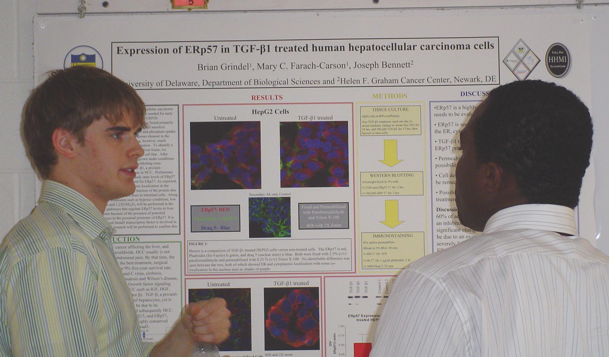

Expression of ERp57 in a TGF-β1-Treated Human Hepatocellular Carcinoma Cell Line

Brian Grindel, Mary C. Farach-Carson, and Joseph Bennett
University
of Delaware, Department of Biological Sciences and Helen F. Graham Cancer Center, Newark, DE

Hepatocellular carcinoma (HCC), a neoplasm of hepatocytes, is the fifth most common cancer worldwide.  Most diagnoses are made when the disease is well advanced, attributing to the poor prognosis of HCC, which carries only a 5% five-year survival rate.  The molecular mechanisms leading to and advancing HCC are not well understood; therefore more information is needed for early identification and potential new therapeutic targets.  ERp57 (GRP58/1,25-(OH)2D3 MARRS), a thiol oxidoreductase found primarily in the endoplasmic reticulum, has been identified in a variety of functions including assembly of the class I MHC complex and phosphate uptake in developing chickens.  Furthermore, there is a conserved Smad3 response element in the promoter region involved in TGF-β signaling.  However, much remains to be elucidated.  To identify a potential role of ERp57 in cell signaling we undertook a study of ERp57 in HEPG2 cells, a human HCC cell line.  After establishing a baseline of ERp57 levels in HEPG2 cells grown under conditions of serum deprivation, experiments with Western immunoblotting were performed. HEPG2 cells were treated with 200 pM TGF-β1, a pro/anti-inflammatory cytokine for hepatocytes whose levels rise in HCC.  Preliminary results indicate that TGF-β1 treatment did increase steady state levels of ERp57 versus controls.  HEPG2 cells also were immunostained for ERp57. As expected, most immunostaining was localized to the ER with some localization in the nucleus.  Along with variable doses of TGF-β1, other treatments such as hypoxic conditions, low glucose, IFN-γ, HGF, TNF-α and 1,25 (OH)2 D3 will be performed in the future to determine other possible pathways for ERp57.  It’s possible the same TGF-β1 conserved Smad3 transcription factor is involved in upregulation of ERp57, but upcoming research will be done to determine this and its role, if any, in active HCC.  [This research was supported and funded by the Howard Hughes Medical Institute and the Center for Translational Cancer Research at the Helen F. Graham Cancer Center.]


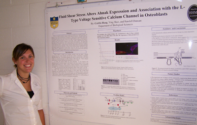
Fluid Shear Stress Alters Ahnak Expression and Association with the L-type Voltage Sensitive Calcium Channel in Osteoblasts

Caitlin Haag, Ying Shao, and Randall Duncan
Department of Biological Science

Strains we encounter through everyday activities are necessary for our skeletal formation and function, yet the cellular mechanisms necessary for this response are unknown.  The earliest reaction to mechanical strain caused by fluid shear in osteoblasts is a rapid increase in intracellular calcium that occurs through activation of the L-type voltage sensitive calcium channel (LVSCC).  This calcium influx is necessary to induce bone formation in vivo (Li et al., 2002).  The actin cytoskeleton also responds to mechanical strain with an increase in formation of stress fibers.  Ahnak, a 700kD protein has recently been found to interact with both the LVSCC and the f-actin of the cytoskeleton.   We postulate that ahnak bridges the cytoskeleton to the LVSCC and that fluid shear will alter the expression of this protein in a time dependent manner.  I found that during fluid shear, ahnak expression decreased in osteoblasts within one hour of the onset of fluid shear.  Ahnak expression returned within 6 hrs suggesting that ahnak can be rapidly degraded and synthesized or is perhaps stored in discrete locations within the cell.  Future studies include cell fragmentation to identify ahnak storage sites within the cell and cytoskeleton disruption with immunofluorescent imaging to determine changes in ahnak localization during application of shear.  This research is funded by NIH and Howard Hughes Medical Institute.


The Role of CIB1 in Cell Adhesion
Tyanna Hadley and Ulhas P. Naik
Department of Biological Sciences

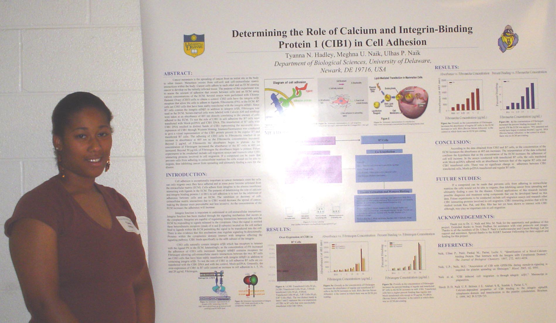
Cell adhesion is vastly important in cancer metastasis since the cells can only migrate once they have adhered and at some point become unbound from the extracellular matrix (ECM). The purpose of this experiment was to assess the amount of adhesion that occurs between cells and an ECM using various concentrations of the ECM. Several assays were performed with Chinese Hamster Ovary (CHO) cells to obtain a control. CHO cells have the integrin 51 receptors that allow the cells to adhere to the ligand Fibronectin in the ECM. B7 cells are CHO cells that have been stably transfected with the integrin IIb3. Since B7 cells contain the integrin IIb3 in addition to integrin 51, Fibrinogen was used as the ECM. Serum-starved cells were labeled with Calcein AM and readings were taken at an absorbance of 485 nm directly correlating to the amount of cells adhered to the ECM. To test the role of CIB1 in cell adhesion the B7 cells were transfected with Mock-pcDNA and CIB1 DNA. The transfection of B7 cells with CIB1 DNA resulted in distinct bands of CIB1 protein between 20 and 30 kDa representing the successful over-expression of CIB1 through Western blotting. Immunofluorescence was conducted to give a visual representation of the CIB1 protein present in the regular B7 and transfected B7 cells. The adhering of CHO cells to Fibronectin resulted in an increase in absorbance at 485 nm as the Fibronectin concentration increased. Beyond 2 g/mL of Fibronectin the absorbance began to plateau. As the concentration of Fibrinogen increased the absorbance of the B7 cells at 485 nm increased. Beyond 50 g/mL of Fibrinogen the absorbance began to plateau. Future experiments to be conducted include cell migration assays and research other CIB interacting proteins involved in cell migration. If a compound can be made that prevents cells from adhering to extracellular matrices the cells would not be able to migrate, thus inhibiting cancer from spreading and ultimately finding a cure for the disease.


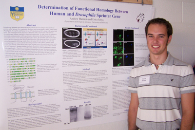
Determination of the Functional Homology Between Human and Drosophila Sprinter

Andrew Harmon and Erica Selva
Department of Biological Sciences

Recently our laboratory has identified a highly conserved novel multipass transmembrane protein, called Sprinter (Srt), required by secretory cells for the release of active Wnt/Wingless (Wg) ligand. Drosophila and Human Srt are 42% identitcal and 60% similar at the amino acid level.  The overall goal of this project is to determine if the conservation of Srt between Drosophila and Humans translates to functional conservation.  In order to test this hypothesis, functional complementation will be performed to demonstrate that the human protein can rescue Drosophila srt loss of function phenotypes. Hence, the immediate goals of the project were to develop constructs that allow for the expression of the human protein in Drosophila with a Haemagglutin (HA) tag, which allows the human Srt to be detected.  A multi-step cloning strategy was used to add the HA coding sequence to the carboxy terminus of the human gene already cloned into pUAST, an in vivo and in vitro Drosophila expression vector, (pUAST-Hsrt-HA).  Despite abundant similarity, the carboxy terminal tails of these proteins differ significantly.  Therefore, a second cloning strategy was used to replace the C-terminal tail of Human Srt with Drosophila sequences containing an HA tag. This chimeric sequence was then cloned into pUAST (pUAST-Hsrt-Dsrt-HA). To determine if the Human Srt protein will be expressed, these constructs will be transfected into Drosophila SL2 and S2R+ cells.  Expression of the protein will then be confirmed by using Western Blot and cell staining analysis using an HA antibody.  Funding for this project was provided the University of Delaware Science and Engineering Scholars Program.


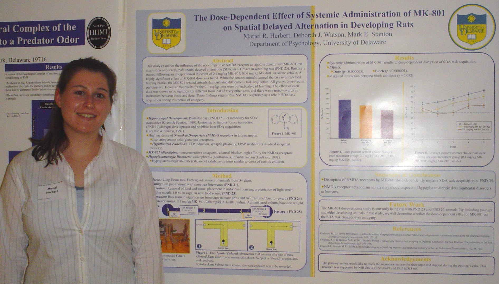
The Dose-Dependent Effect of Systemic Administration of MK-801 on Spatial Delayed Alternation in Developing Rats

Mariel R. Herbert
, Deborah J. Watson, and Mark E. Stanton
Department of Psychology

Spatial working memory has been shown to rely upon hippocampal development between postnatal day (PND) 15 and 21 (Green & Stanton, 1989). When development is interrupted by lesioning or fimbria-fornix transection on PND 10, juvenile rats (PND 23) are unable to successfully learn a discrete-trials spatial delayed alternation (SDA) task (Freeman & Stanton, 1991). Spatial working memory may also be disrupted later in development via N-methyl-D-aspartate (NMDA) receptor channel blockers. It is possible that synaptic plasticity is dependent upon glutamate receptor function. This study examines the influence of the noncompetitive NMDA receptor antagonist dizocilpine (MK-801) on SDA acquisition in a T-maze task in weanling rats (PND 25) following an interperitoneal injection of 0.1 mg/kg MK-801, 0.06 mg/kg MK-801, or saline vehicle. A highly significant effect was found for dose. While the control animals learned the task over repeated training, the MK-801-treated animals demonstrated difficulty in task acquisition. All groups improved in performance. However, the results for the 0.1 mg/kg dose were not indicative of learning. The effect of each dose was shown to be significantly different from that of every other dose, and a trend towards an interaction between block and dose was also discovered. These findings suggest that the impairment of NMDA receptors by MK-801 interferes with SDA task acquisition during ontogeny. Supported by NIH R01 AA014288-01 and PO1 HD35466. 


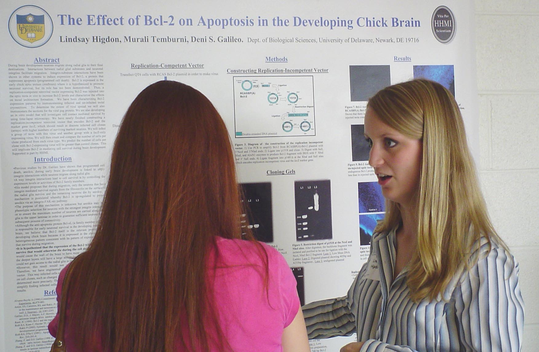
The Effect of Bcl-2 on Apoptosis in the Developing Chick Embryo Brain

Lindsay Higdon and Deni S. Galileo
Department of Biological Sciences

During brain development neurons migrate along radial glia to their final destinations.  Interactions between radial glial substrates and neuronal integrins facilitate migration.  Integrin-substrate interactions have been shown in other systems to induce expression of Bcl-2, a protein that suppresses apoptosis (programmed cell death).  Bcl-2 is expressed in early chick optic tectum (midbrain) where it is hypothesized to promote neuronal survival, but its role has not been demonstrated.  Thus, a replication- competent retroviral vector expressing Bcl-2 was injected into the optic tecta in vivo to increase Bcl-2 levels and characterize the effects on tectal architecture formation.  We have been characterizing Bcl-2 expression patterns by immunostaining infected and uninfected tectal cryosections.  To determine the extent of viral spread we will also immunostain the sections for the viral gag protein. We are also developing an in vitro model that will investigate cell contact mediated survival by using time-lapse microscopy. We have nearly finished constructing a replication-incompetent retroviral vector that encodes Bcl-2 and the marker gene lacZ, which should result in discrete infected cell clones (arrays) with higher numbers of surviving marked neurons. We will infect a group of tecta with this virus and another group with a lacZ-only expressing virus. We will then count and compare the number of cells per clone produced from each virus type. We predict the number of cells per clone with Bcl-2-expressing virus will be greater than control clones.  This will implicate Bcl-2 in mediating cell survival during brain development.  Supported in part by HHMI.


The Relationship Between Water Quality and Horse Activity near Ribbed Mussel Beds
along Assateague Island National Seashore, Maryland

Janaire L. Hughes
, Mary S. Lambert, and Gulnihal Ozbay
Department of Agriculture and Natural Resources

Delaware
State University, Dover, Delaware

Assateague Island National Seashore, MD supports approximately 150 feral horses (Equus caballus).  The objective of this study was to determine the relationship between these horses and water quality near ribbed mussel (Geukensia demissa) beds. Five study sites were selected south to north, containing ribbed mussels and varying horse activity.  At each site, salinity, temperature, total ammonia nitrogen, and nitrite were measured. Total bacteria counts and Vibrionaceae levels were determined using Colony Overlay Procedure for Peptidases.  There was a noticeable increase in both bacteria and Vibrionaceae levels at all sites from June to July 2006. Total bacteria levels at site 23.4 increased from 17,400 to 183,780 bacteria/g of mussel, while Vibrionaceae levels increased from 5,300 to 103,570 Vibrionaceae/g of mussel.  Total bacteria levels at site 18.9 increased from 74,000 to 199,330 bacteria/g of mussel, while Vibrionaceae levels increased from 22,330 to 110,340 Vibrionaceae/g of mussel.  At site 17.4, bacteria levels increased from 166,930 to 365,530 bacteria/g of mussel, while Vibrionaceae levels increased from 77,860 to 282,680 Vibrionaceae/g of mussel.  At the south bridge site, bacteria levels increased from 44,560 to 205,830 bacteria/g of mussel, while Vibrionaceae levels increased from 25,560 to 169,500 Vibrionaceae/g of mussel.  Total bacteria levels at the north bridge site increased from 572,200 to 1,353,330 bacteria/g of mussel, while Vibrionaceae levels increased from 265,030 to 635,000 Vibrionaceae/g of mussel. Future samples should be taken from the northern end of the island (presently closed), which has decreased horse activity to determine if horse activity influences the increased bacteria levels or if other factors are involved. Funding for this research was provided by USDA/CSREES Grant 2004 -38820-15154 awarded to Dr. Gulnihal Ozbay and EPSCoR- supported by NSF EPSCoR Grant No. EPS-0447610.


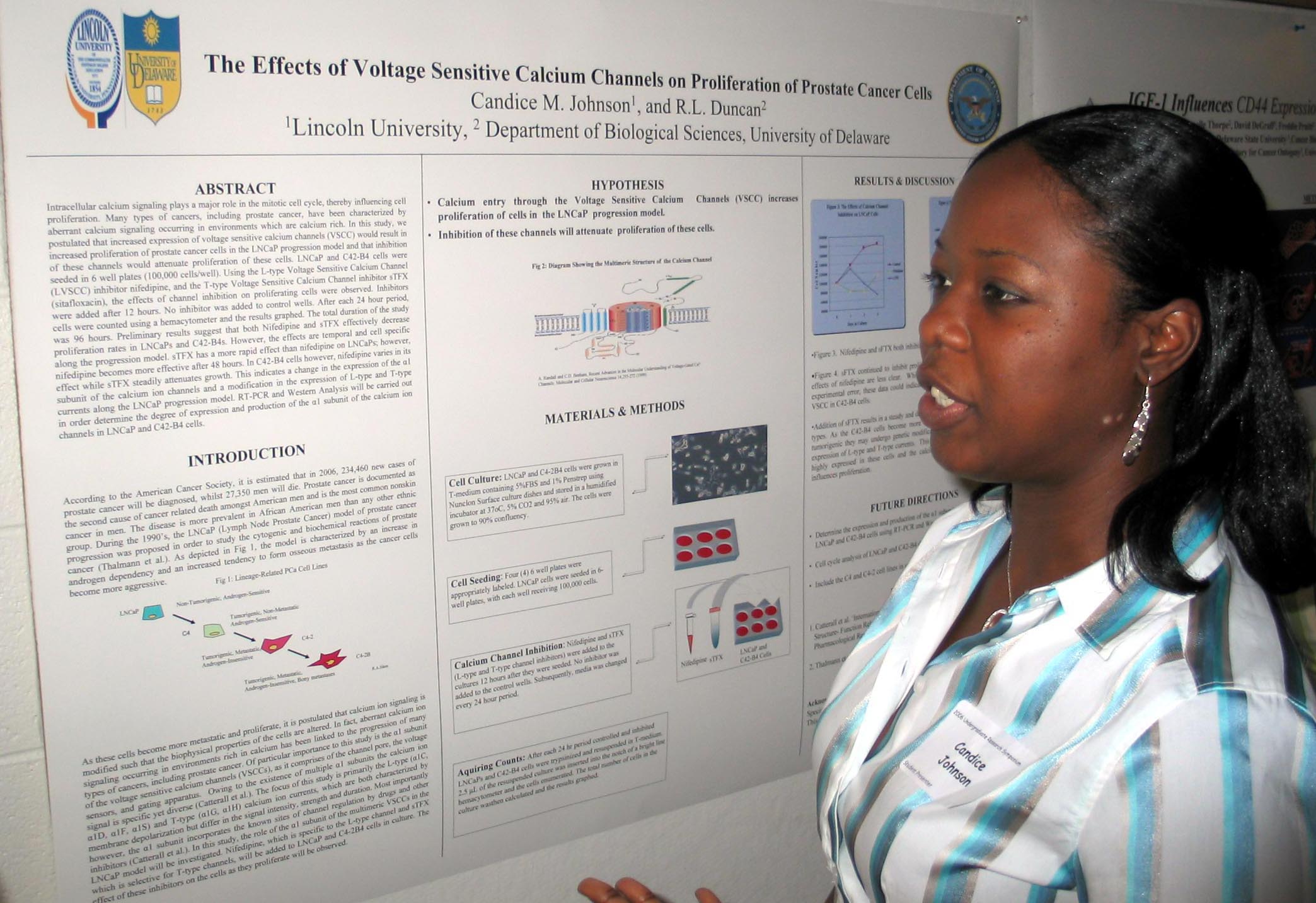
The Effects of Voltage Sensitive Calcium Channels on Proliferation of Prostate Cancer Cells.

Candice M. Johnson
1, and Randall L. Duncan2
1
Lincoln University, 2 Department of Biological Sciences, University of Delaware

Intracellular calcium signaling plays a major role in the mitotic cell cycle, thereby influencing cell proliferation. Many types of cancers, including prostate cancer, have been characterized by aberrant calcium signaling occurring in environments which are calcium rich. In this study, we postulated that increased expression of voltage sensitive calcium channels (VSCC) would result in increased proliferation of prostate cancer cells in the LNCaP progression model and that inhibition of these channels would attenuate proliferation of these cells. LNCaP and C42-B4 cells were seeded in 6 well plates (100,000 cells/well). Using the L-type Voltage Sensitive Calcium Channel (L-VSCC) inhibitor nifedipine, and the T-type Voltage Sensitive Calcium Channel inhibitor sTFX (sitafloxacin), the effects of channel inhibition on proliferating cells were observed. Inhibitors were added after 12 hours. No inhibitor was added to control wells. After each 24 hour period, cells were counted using a hemacytometer and the results graphed. The total duration of the study was 96 hours. Preliminary results suggest that both Nifedipine and sTFX effectively decrease proliferation rates in LNCaPs and C42-B4s. However, the effects are temporal and cell specific along the progression model. sTFX has a more rapid effect than nifedipine on LNCaPs; however, nifedipine becomes more effective after 48 hours. In C42-B4 cells however, nifedipine varies in its effect whilst sTFX steadily attenuates growth. This indicates a change in the expression of the α1 subunit of the calcium ion channels and a modification in the expression of L-type and T-type currents along the LNCaP progression model. RT-PCR and Western Analysis will be carried out in order determine the degree of expression and production of the α1 subunit of the calcium ion channels in LNCaP and C42-B4 cells. This project was funded by the Department of Defense.


Identification of Nodulation Mutants in an EMS-mutagenized Population of Medicago truncatula

Laura D. Johnson1, 2*, Liana T. Burghardt2, 4, Heather Danysh2, 3, David H. McNear2,3, and D. Janine Sherrier1, 2, 3
1Department of Biological Sciences, University of Delaware, 2Delaware Biotechnology Institute, University of Delaware, 3Department of Plant and Soil Sciences, University of Delaware, and 4 Department of Biological Sciences, Carleton College

Medicago truncatula, a leguminous plant, forms a symbiotic relationship with the soil bacterium, Sinorhizobium meliloti. S. meliloti induces nodule formation on the roots of M. truncatula; nodules are organs in which atmospheric nitrogen is converted into a chemical form that is usable by the host plant. In this study, my objective was to isolate plants that were altered in nodule formation or function from an ethyl methane sulfonate (EMS) - mutagenized population. Two hundred and twenty five plants from the EMS population and thirty- seven wild- type were grown aeroponically for 28 days and were screened for nodule formation and nodule development.  Plants were scored according to their nodule and root phenotypes, including nodule number, nodule size, nodule color, and root length. The most significant mutant phenotypes consisted of white, potentially non-fixing nodules (nod+ fix-) and plants that lacked nodule formation altogether (nod-). Initial screenings identified 17 putative mutants. Out of the 17 nodulation mutants, 7 have white nodules and 10 have no nodules. With the exception of the nodule phenotype, the mutant plants appeared to be healthy with morphology similar to wild- type control plants. The mutant plants were comprised of long shoots, green leaves, which varied in size, an extensive lateral root system, an intact main root, and, in general, a length greater than 20 cm. These 17 were planted in pots for seed collection and future phenotypic characterization of their progeny. Supported by an undergraduate research scholarship funded by the Delaware Biotechnology Institute’s NSF EPSCoR Grant No. EPS-0447610.


Broiler Stress and Welfare: Physiological and Behavioral Impacts of Elevated Ammonia

Tanveer Kahn and Sabrina M. Brougher
Department of Biology, Delaware State University, Dover DE.

An historic problem faced by the poultry industry is the negative impact of elevated ammonia (NH3) bird quality and welfare. Research on the impact of NH3 on relevant endpoints (e.g. pulmonary quality, stress and behavior) is limited. Mammalian pulmonary surfactant proteins (SP) aid airway inflammation and modulate immune response, and avian SP likely function similarly. We hypothesize that elevated NH3 may compromise the efficacy of the avian pulmonary system via disruption of the SP profile, and that this reduction in respiratory capacity will manifest as elevated stress and increased fear response in broilers.  The objective of the current project is to determine if elevated NH3 alters broiler physiological development in terms of growth, fear response, measured as the duration of tonic immobility (TI), stress, measured by both plasma corticosterone (CORT) and the degree of fluctuating asymmetry (FA) in bilateral traits, and pulmonary SP expression.  Mixed sex broiler hatchlings will be randomly assigned to 12 positive pressure poultry isolation units (30 birds/isolator) with ad libitum feed and water and a standard commercial lighting regime.  Six isolators will remain under low (L) NH3 and the remaining 6 will receive high (H) NH3 of 25 ppm continuous exposure. Across 6 wks, a weekly subset of 5 birds / isolator will be weighed, culled and lungs retained for SP analysis (Western immunoblot).  At week 6, wing vein blood will be collected for plasma CORT analysis (ELISA), and tarsometatarsal measurements will be recorded for FA analysis. Funded by Delaware EPSCoR (under NSF Grant No. EPS-0408729).


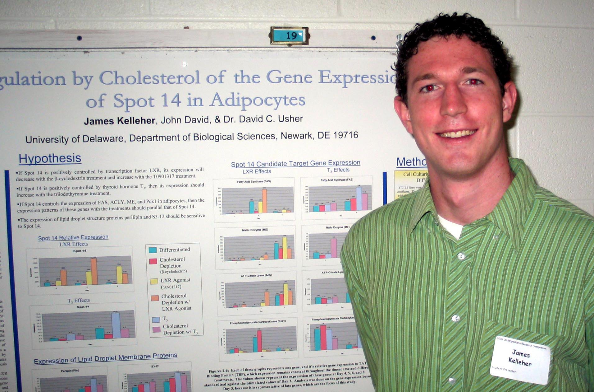
Regulation by Cholesterol of the Gene Expression of Spot 14 in Adipocytes

James Kelleher, John David, and David Usher
Department of Biological Sciences

Thyroid hormone responsive Spot 14 (THRSP) is a transcription factor localized to the liver and adipocytes.  In addition to sensing triiodo-L-thyronine (T3) and glucose levels, previous studies have indicated the expression of Spot 14 is regulated by liver X receptor (LXR), a transcription factor activated by cholesterol.  The known target genes of Spot 14 are specific to lipogenesis, suggesting it as a control point in the accumulation of lipids.  In adipocytes, however, the regulation and function of Spot 14 have yet to be investigated.  In this study, 3T3 L1 mouse adipocytes depleted of cholesterol and supplemented with an LXR agonist and/or T3 were used to examine the regulation of Spot 14.  Gene expression of these treated cells was measured through quantitative PCR (QPCR) for Spot 14 and its hypothesized target genes.  In what appears to be through independent mechanisms, the expression of Spot 14 increased with both the additions of T3 and the LXR agonist, whereas the cholesterol depletion had the opposite effect showing a decrease in the mRNA.  The proposed target genes fatty acid synthase (FAS), ATP-citrate lyase (Acly), and phosphoenolpyruvate carboxykinase (Pck1) demonstrated a similar, yet T3 dominant response.  Of particular interest were the results for perilipin, a protein found on the surface of large lipid droplets.  The expression of this gene also exhibited a cholesterol dependent response similar to that of Spot 14.  The results from this study provide further indication of the cholesterol-mediated role of Spot 14 in the storage of lipids. This research was funded in part by the Howard Hughes Medical Institute Undergraduate Science Education program.


Tumor Necrosis Factor Alpha (TNF-α) Regulation of Reticulocalbin (RCN-1) Cell Surface Expression
and Capillary-like Formation in Bone Marrow Endothelial Cells (BMECs)

Andrew Koemeter-Cox
1, Rachel Oren2 , Jill Lynch1, Mary C. Farach-Carson1, and Carlton R. Cooper1
1
Department of Biological Sciences, University of Delaware, Newark, DE and
2Department of Allied Health-Histotechnology, Delaware Technical Community College, Wilmington DE

The goal of this project is to determine the effect that Tumor Necrosis Factor Alpha (TNF- α) has on Bone Marrow Endothelial Cells (BMECs).  Specifically, Reticulocalbin (RCN-1) surface expression and capillary-like formation of the BMECs were examined.  TNF-α is a pro-inflammatory cytokine known to be involved in inflammation and angiogenesis, while RCN-1 is a recently discovered surface protein whose function has not yet been reported.  It is hypothesized that RCN-1 levels on the cell surface will be increased by TNF-α, while its effect on angiogenesis of BMECs is unknown.  To determine RCN-1 surface expression, BMECs were given three different treatments of TNF-α.  One group was given none, another group was treated for four hours with TNF-α and the third group was treated for twenty-four hours.  The cells were treated with an RCN-1 anti-body, and then treated with a secondary fluorescent anti-body.  Analysis with Fluorescence Activated Cell Sorting (FACS) shows that the levels of RCN-1 were noticeably increased on the BMECs treated with TNF-α.  To determine the effect of TNF-α on capillary-like formation, BMECs were left untreated and treated with TNF-α for four hours.  Each group was then grown on Matrigel® for twenty-four hours in plain media and media containing TNF-α.  The cells were examined after the growth period, and only the BMECs that never were exposed to TNF-α showed significant capillary-like formation.  This, along with the previously mentioned RCN-1 levels, suggests that up-regulation of RCN-1 on the BMEC cell surface by TNF-α results in a drastic decrease in angiogenesis.  Funding for this project was provided by INBRE.


Biology Abstracts for students with last names starting with L-Z.

Links: Summer 2006 Undergraduate Research Symposium, Symposium Abstracts from other Colleges and Departments,
Undergraduate Research Summer Enrichment ProgramUnversity of Delaware Undergraduate Research Program, Howard Hughes Undergraduate Program.
Created  1 August 2006. Last up dated 16 August 2006 by Hal White
Copyright 2006, University of Delaware