 |
JAZZ IT UP AT EXPERIMENTAL BIOLOGY MEETINGS IN NEW ORLEANS |
 |
|
JAZZ IT UP AT EXPERIMENTAL BIOLOGY MEETINGS IN NEW ORLEANS |
|
 |
New Orleans, the home of paddle wheelers, crawdads, and Bourbon Street, was host of the 2002 Experimental Biology Meetings where seven Unversity of Delaware students presented their research at the Undergraduate Research Poster Session sponsored by the American Society for Biochemistry and Molecular Biology (ASBMB), April 20 to April 24. In addition to HHMI sponsorship, each of the students received a travel supplement from ASBMB. |
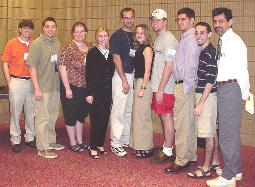
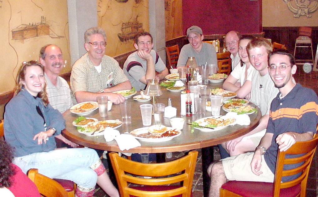
The University of Delaware group included three faculty and seven undergraduates.
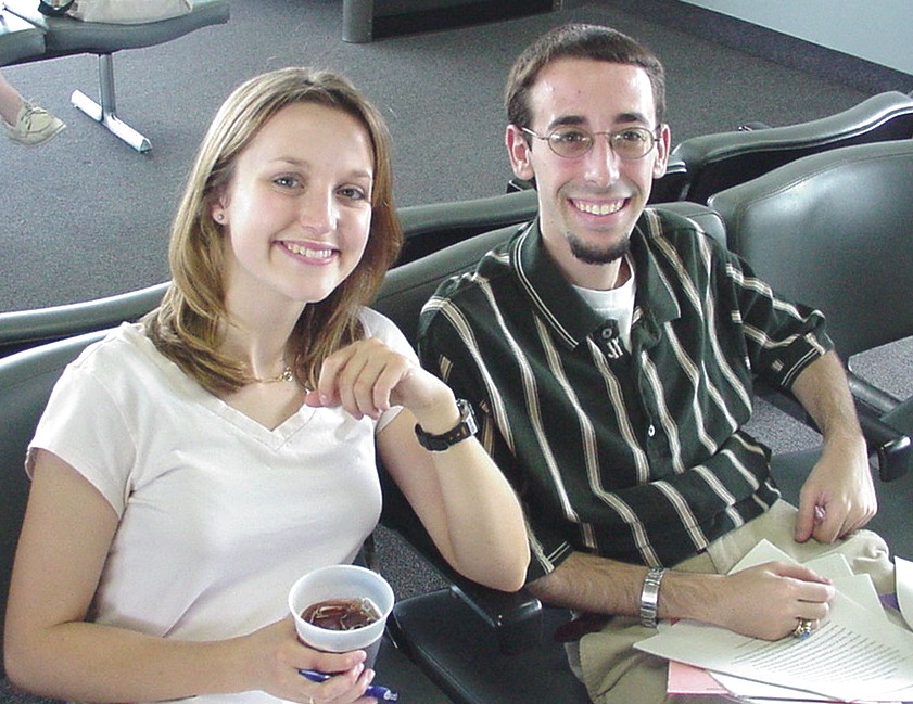
| At River's Edge Restaurant
from left to right Suzanne Biehn (Sr. Biological Sciences) Prof. Gregory Stephens (Biological Sci.) Prof. Hal White (Chem. and Biochem.) Eugene Antipov (Sr. Chem Eng & Biochem) Kevin O'Hayer (Sr. Biological Sciences) Prof. David Usher (Biological Sci.) Courtney Smith (Sr. Medical Technology) Michael Usher (Sr. Biological Sciences) Justin DiAngelo (Sr. Biotechnology) (Alice Wong Jr. Med. Tech. not shown) |
 |
 |
Suzanne Biehn and Justin DiAngelo received First Place Awards in the ASBMB Undergraduate Poster Competition sponsored by the Portland Press/Biochemical Journal. |
|
|
 |
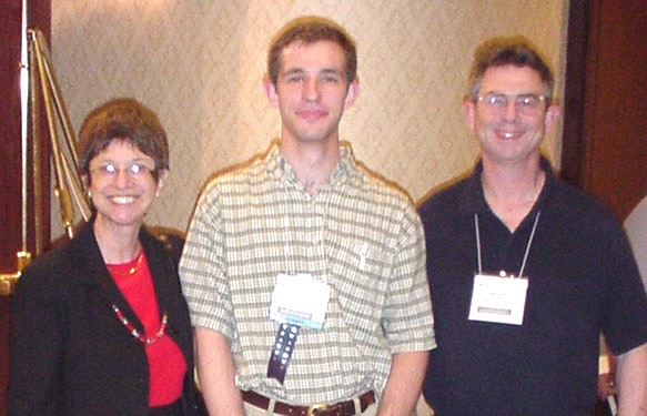 |
Eugene Antipov poses with Judy and Don Voet, authors of the Biochemistry textbook he used in CHEM-642. The Voets, also editors of Biochemistry and Molecular Biology Education, were among the 20 judges for the Undergraduate Poster Competition. |
|
|
|
|
|
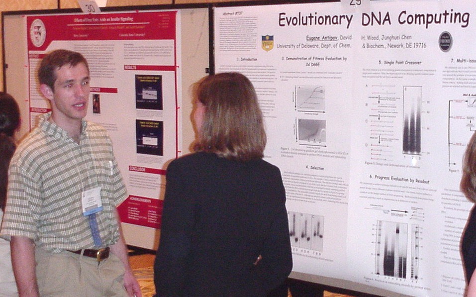
Eugene Antipov, David Harlan Wood, and Junghuei Chen
|
This paper is the first attempt to demonstrate DNA implementation of a class of Evolutionary Computation algorithms, an established paradigm for conventional computers. In silico, evolutionary computation is often used for solving problems involving many interacting variables such as timetable construction, machine learning, and graph drawing layout. Evolutionary computation seems particularly well suited to DNA implementation because of its alleged robustness in the presence of errors and its ability to exploit massive parallelism and memory inherent at the molecular level. Here we present the first DNA implementation of evolutionary computation and compare its performance with in silico simulations. It is demonstrated using selection by fitness, and breeding via crossover and mutation. All aspects of our DNA implementation of evolutionary computation are tested using a simple example problem. We limit ourselves to a special class of problems where we completely forgo all arithmetic, relying instead on statistical comparisons. However, when applied to our class of problems, conventional computers cannot match our parallel processing power and memory capacity. |
|
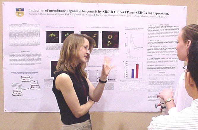
biogenesis by Ca 2+ -ATPase (SERCA1a) expression. Suzanne E. Biehn, Jeremy M. Lyons, Kirk J. Czymmek, and Norman J. Karin.
|
The mechanisms by which sarcoplasmic reticulum (SR) arises during skeletal myogenesis are unknown. SR has been proposed to emerge from the endoplasmic reticulum (ER) and the biosynthesis of SR/ER Ca 2+ -ATPase, SERCA1a, is among the earliest events of skeletal myogenesis. We hypothesize that SERCA1a expression is a stimulus for organelle membrane biosynthesis. Overexpression of avian SERCA1a in transfected mouse Ltk - fibroblasts elicited compact masses of intracellular membrane (plaques) that were enriched in SERCA1a protein. Unfixed cells transfected with cDNA encoding a SERCA1a/Green Fluorescent Protein fusion protein demonstrated that membrane plaques were not fixation-induced artifacts. Immunofluorescent labeling indicated co-localization of SERCA1a with the ER marker calreticulin, although western blots showed that the level of endogenous ER protein expression was not altered in SERCA1a-transfected cells. These data suggest that SERCA1a expression triggered the generation of specialized membrane structures that are contiguous with the ER and contain ER proteins. Supported by the University of Delaware Research Foundation and Pfizer, Inc. |
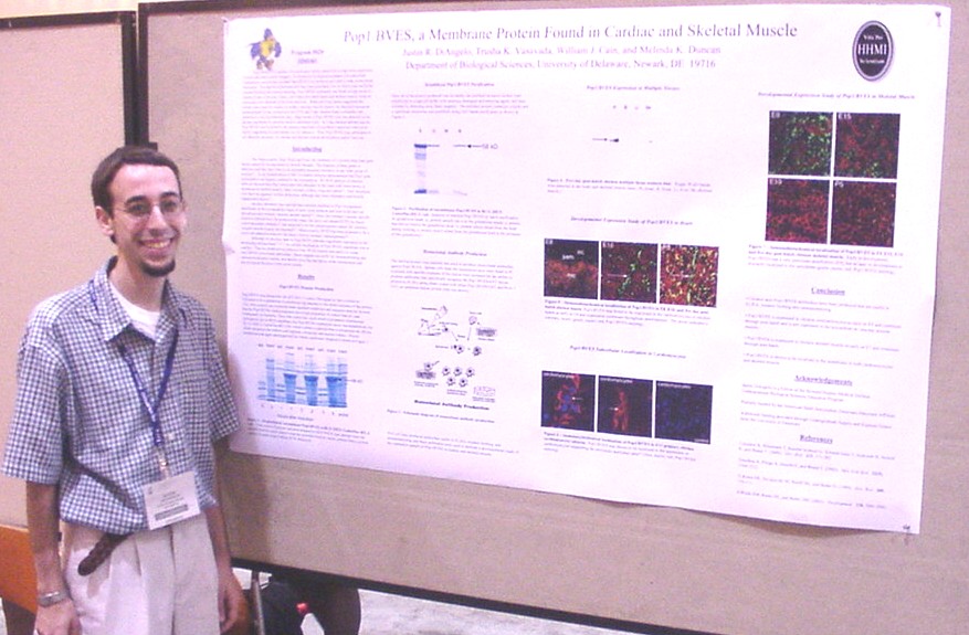
| Pop1/BVES is a member of a novel gene family named for its high level expression in heart and other muscle lineages. To determine its expression pattern and subcellular localization, recombinant chicken Pop1/BVES was produced and used to make monoclonal antibodies. Ten specific hybridoma cell lines were generated, two of which were useful for western blotting and immunostaining. Pop1/BVES expression was found at high levels in chicken 8-day embryonic hearts, and 5-day post-hatch hearts and skeletal muscle while no expression was detected in the brain and liver. While previous reports suggested this protein was a specific marker of cardiac vascular smooth muscle, we detected expression predominantly in the cardiomyocytes of E8 and 5-day chicken hearts consistent with published in situ hybridization data. High levels of Pop1/BVES were also detected at the plasma membrane in cultured chicken cardiomyocytes. In 5-day chicken skeletal muscle, Pop1/BVES was localized to the plasma membrane of myofibers consistent with recent reports suggesting its participation in cell adhesion. Thus, Pop1/BVES may participate in cell adhesion necessary for cardiac and skeletal muscle development and/or function. |
Found in Cardiac and Skeletal Muscle Justin Robert DiAngelo, Trusha Kirit Vasavada, William Cain, and Melinda Kay Duncan
|
|
inhibitory to DNA binding in SV-40 Large T-Antigen Kevin Mitchell O'Hayer, Rebecca Parsons, Rupa Roy, and Daniel T. Simmons 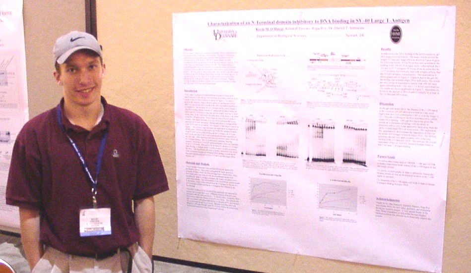 |
SV-40 Large T-Antigen is a multifunctional protein, which is responsible for the transformation of cells upon viral infection. The region of the protein which this experiment deals with, is the DNA binding domain at the N-Terminus of the protein. The DNA binding domain has been elucidated and characterized extensively by the use of deletion mutants. It lies in the region corresponding to residues 131-259. This inquiry utilized N-terminal fragments created in a baculovirus expression system and purified utilizing an intein tag that was cleaved from the protein in the purification process. Once the proteins were purified, they were utilized in gel shift assays to determine the DNA binding characteristics of each individual fragment. It was found that the 1-109 region of the protein lowered the DNA binding activity in the gel shift assays of the proteins involved. Proteins missing this region were better suited to DNA binding of both the 112 base pair full origin and 64 base pair minimal origin. |
|
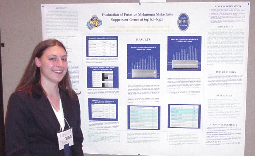
Melanoma Metastasis Suppressor Genes at 6q16.3-6q23 Courtney Smith, Carrie Paquette-Straub, and Mary E. Miele
|
Previous studies show that introduction of a human chromosome 6 (chr6) causes suppression of metastasis in a nude mouse model. Additional studies localize the metastasis suppressor locus to 6q16.3-q23. Evidence of this was provided by assaying metastatic cells containing an added chr6 lacking 6q16.3-23.3 (6qdel). The specific genes responsible for metastasis suppression are unknown. These genes can be found by comparing the expression of gene(s) in nonmetastatic cells (with an added chr6) to metastatic 6qdel containing cells. To test differential gene expression, various metastatic and non-metastatic hybrid cell lines originating from the C8161 parent line were studied for expression of two putative metastasis- suppressor genes that localize to 6qdel. These two genes, Connexin 43 and Vitamin D3 interacting protein, were analyzed using RT-PCR. Total RNA was collected from 15 cell lines. Results indicate multiple gene interactions are involved in regulating metastasis suppression. To identify additional genes involved, microarray experiments will be performed. Research support: Howard Hughes Medical Institute (CS), UD Undergraduate Research Program, and 1R15CA88876-01 (MEM). |
|
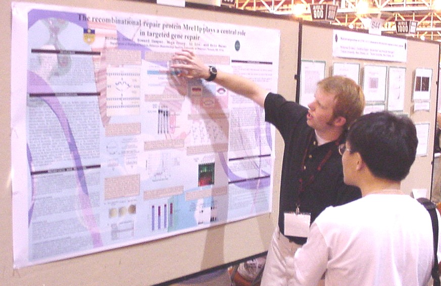
Mre11p, plays a central role in targeted gene repair Michael Goodwin Usher, Howard Gamper, Miya Drury, Li Liu, and Eric Kmiec
|
Targeted Gene Repair is
a method that utilizes oligonucleotide vectors to illicit sequence specific
mutagenesis. This process is initiated by recombinase-mediated pairing
events in which the vector hybridizes to a target strand, creating a displacement
loop (D-loop) intermediate. The gene repair event is directed by the oligonucleotide,
which serves as the template for the transfer of base pair information
to the target site. Finally, the joint is resolved by endolytic cleavage
and strand passage, leaving an intact, converted nucleotide. The mechanism
of gene repair is being elucidated with a practical fallout of understanding
the factors that influence repair frequency. Using yeast as a model system, we have identified several proteins that play essential roles in the process, including Mre11p and Rad50p. Biochemical analysis shows that Mre11p acts to resolve the D-loop by specific nucleolytic cleavage, a reaction in which the specificity of cut sites is directed by Rad50p. These data suggest that proteins involved in the processing of the D-loop resolve the intermediate and increase the probability that converted nucleotides are maintained as a result of the gene repair process. |
|
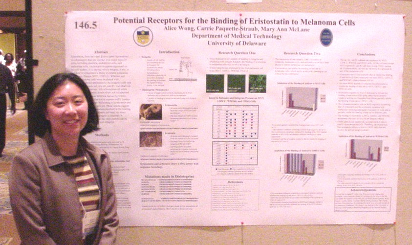
of Eristostatin to Melanoma Cells Alice Wong, Carrie Paquette-Straub, and Mary Ann McLane
|
Eristostatin, from the viper
Eristocophis macmahoni, is a disintegrin that can interact withnmany types
of cells, including platelets, endothelial cells, and melanoma cells, via
integrin receptors expressed on the cell surface. It is unclear which integrin,
if any, is involved in eristostatin's ability to inhibit melanoma metastasis.
Human MV3, 1205LU, WM164, and C8161 melanoma cells were incubated with
fluorescent-labeled antibodies to the integrin alpha-v beta-3 and the integrin
subunits alpha-2, -4, -6, and -1, and observed by confocal microscopy.
All cells express alpha 2 while none express detectable levels of alpha-6.
Alpha-4 is expressed by 1205LU, MV3, and WM164, but not by C8161. MV3 is
the only cell line not to express alpha-v beta-3. Studies were also done
to assess the binding of eristostatin and a panel of eristostatin mutations
to anti-alpha 4. These results suggest that, while the critical residues
involved in the binding of eristostatin to the melanoma cells vary, an
intact RGD sequence in the
disintegrin is essential. It remains to be seen whether the same pattern can be determined with the other cell integrins. |