


Imaging capability
UD to house state's first functional MRI instrument


9:59 a.m., April 24, 2015--When a doctor examines an injured athlete for a possible torn ligament, she wants to get a detailed look at the structure of that ligament, and magnetic resonance imaging (MRI) can provide exactly what she needs.
But when a researcher is studying how the human brain works, a standard MRI is often not enough.
Research Stories

Chronic wounds
Prof. Heck's legacy
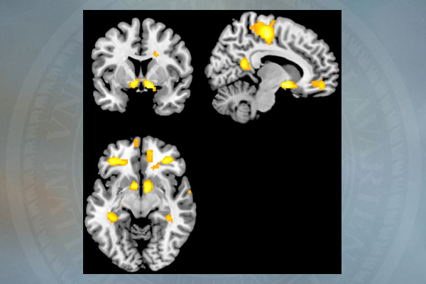
“You can’t tell much about the brain by its structure alone. You need to be able to see how it functions,” said Robert Simons, professor and chair of the University of Delaware’s Department of Psychological and Brain Sciences. “When you have an MRI that shows function, you can see parts of the brain ‘light up’ when a task is performed, and that shows you which structures and neural networks the brain is using while engaged in the task at hand.”
A specialized type of MRI, called fMRI for “functional,” is needed for that type of imaging, Simons said. A research-quality fMRI offers other features as well, including a stronger magnet that can provide more detailed pictures than a standard medical image and will also benefit scientists who need higher-resolution images of the structures that they investigate.
And soon — for the first time on campus and in the state of Delaware — UD will have one of these instruments.
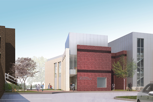
Construction began in mid-March on a two-story extension to the Life Sciences Research Facility on Delaware Avenue. When completed in early 2016, the 11,800-square-foot addition will provide space for an fMRI machine, which will be delivered and lowered into the building’s first floor by crane, probably in January. Plans call for construction to be completed in February and for staff members to move in shortly after.
The addition, known as a multimodal imaging center, will also have conference and office space, facilities for visitors and patients, flexible research space for satellite experiments before and after imaging and space for researchers to house smaller imaging instruments. Some of the other instruments will be in place soon after construction, while others may be acquired over time.
But the large, research-quality fMRI has been ordered and is the priority, said Simons, who with Tatyana Polenova, professor of chemistry and biochemistry, led the faculty task force to plan the imaging facility.
“Before this building, we’ve had no way of doing any of this kind of [cutting-edge brain] research,” he said. “And in competing with universities in Maryland, Pennsylvania and New Jersey, for example, that do have research MRIs, we were clearly at a disadvantage. … President Harker supported this project because he recognized that this kind of imaging capability can no longer be limited to universities with medical schools; there’s a demand for it in all kinds of disciplines.”
In developing proposals for the new facility, Simons and others on the task force held meetings across campus and found researchers in many departments and colleges who were either already using MRIs in their work or were hoping to do so. Faculty members relying on this kind of imaging, Simons said, have been spending time traveling out of state and paying high hourly rates to use research MRIs at other institutions.
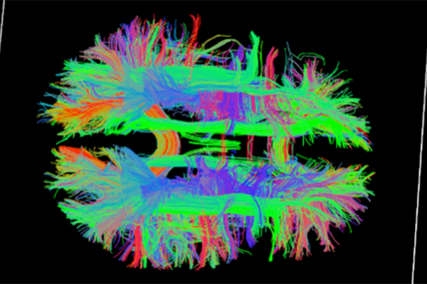
Those instruments, like standard MRIs used routinely for medical diagnosis of a number of conditions, use a large magnet and sequences of radio waves to get a picture of organs and structures in the body, as well as to analyze inanimate objects.
In addition to functional images of the brain, UD researchers make use of MRIs to study such things as tumors in chickens, damage to discs in the spine, bone development in children and the structure of materials. When the facility opens next year, Simons said he expects it to be in demand by researchers in physical therapy, mechanical engineering, materials science, animal science and numerous other fields.
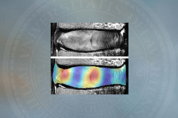
“This type of imaging is critically important, not just for psychology but also for biomedical engineering and health sciences,” said Dawn Elliott, director of UD’s biomedical engineering program. “Having this here will be a big benefit to my research and to many others.”
Elliott, who studies the effects of aging on the discs in the spine that become compressed over time, currently uses MRI to evaluate these intervertebral discs in human and goats. She has been transporting the goats from the University of Pennsylvania’s New Bolton Center School of Veterinary Medicine to center city Philadelphia to scan them in an MRI at the university’s main campus.
“It’s very expensive and time-consuming, and [the travel] is stressful for the animals,” Elliott said. After the scan, the goats are returned, unharmed, to New Bolton, which is near Kennett Square, Pennsylvania, about 15 miles from the UD campus.
Another UD researcher who uses fMRI technology at other locations is Timothy Vickery, assistant professor of psychological and brain sciences, who studies the neural basis of reward learning. He uses fMRI to assess brain activity and connectivity among brain regions during reward-driven learning, to better understand how decisions are made on the basis of past experience.
“I’m very, very excited about the MRI facility, and I think it will be an important research and recruitment tool for Psychological and Brain Sciences as well as many other departments and colleges throughout the University,” Vickery said. “I am sure that it will have a large impact on undergraduate education as well.”
Charlie Riordan, deputy provost for research and scholarship, said support for the imaging facility has been “overwhelming” among faculty members.
“Establishing this new core facility is essential in allowing faculty in many disciplines to examine the internal structure of materials and living things,” Riordan said. “This capability can potentially benefit the work of scholars in all of our colleges. This initiative aligns well with priority recommendations in UD’s new strategic plan aimed at excellence in multidisciplinary research and scholarship.”
The multimodal imaging facility project is supported by the University, the Unidel Foundation and the colleges of Arts and Sciences, Health Sciences and Engineering. The building was designed by MGA Architects and is being built by Bancroft Construction Co. The project manager is Marcia Hutton, UD Facilities Planning and Construction.
Article by Ann Manser
MRI images courtesy of Robert Simons and Dawn Elliott