Abstracts from the College of Agriculture and Marine Studies
Undergraduate Summer Research Symposium August 11, 2004

Ordered alphabetically by student's last name


Colonization of Intertidal Habitats by Invasive Species: Effects of Riprap Emplacement
Abigail M. Bradley1 and Douglas C. Miller2
1
Department of Biological Sciences, 2Graduate College of Marine Studies


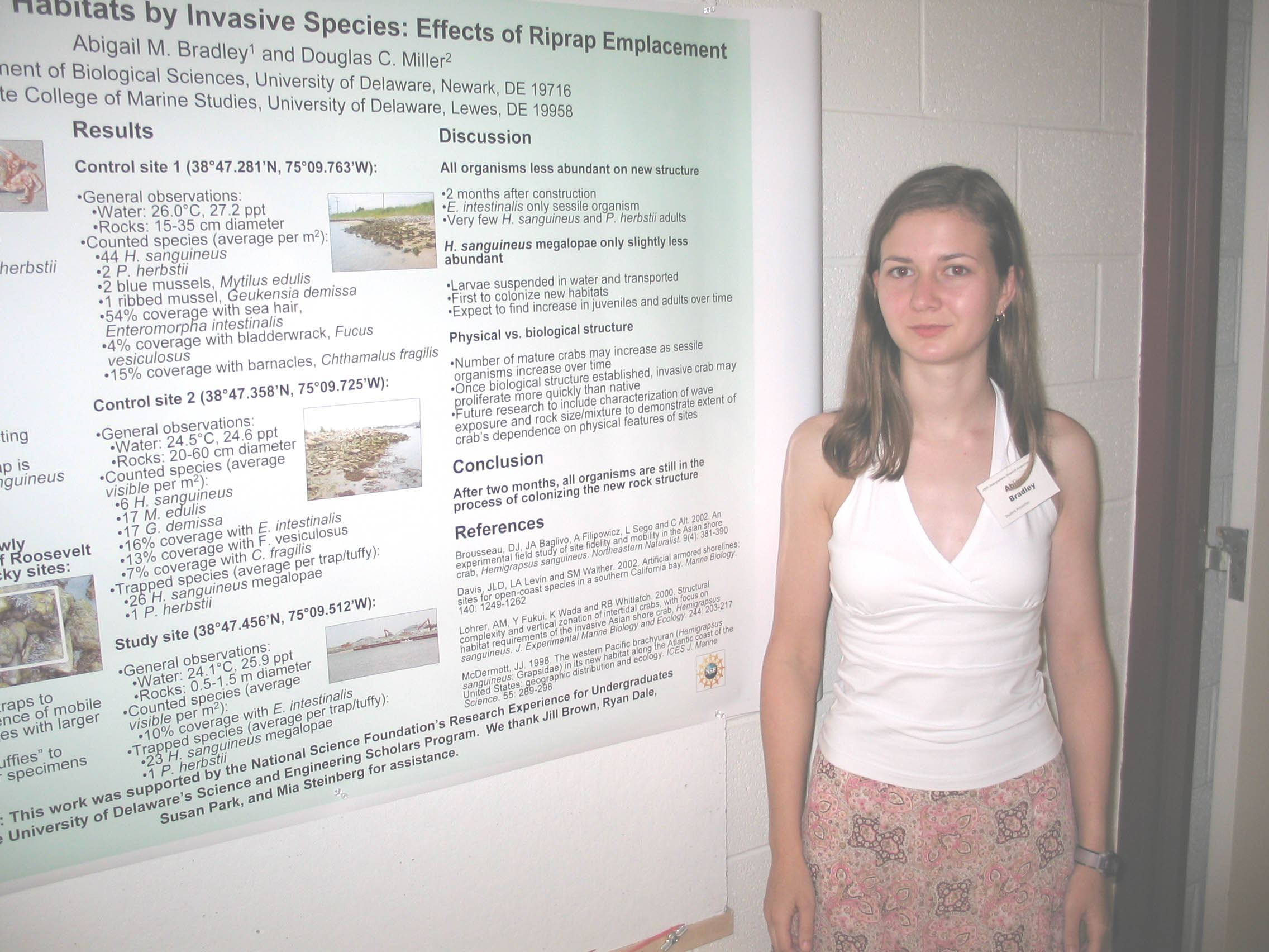
The invasive Asian shore crab, Hemigrapsus sanguineus, was first found in southern New Jersey in 1988, most likely introduced by ballast water discharge.  It has since invaded rocky intertidal habitats of the Atlantic coast from Maine to North Carolina.  Artificial hardening of the shoreline to combat erosion may inadvertently facilitate the spread of invasive species.  Newly emplaced riprap on the south side of Roosevelt Inlet, Delaware Bay, allowed us to monitor colonization with particular emphasis on H. sanguineus.  We compared intertidal communities to those found at nearby rocky sites using quadrat counts, photographs, and minnow traps.  Common organisms included H. sanguineus, mud crabs, amphipods, nereids, mud snails, ribbed and blue mussels, oysters, barnacles, hydroids, bladderwrack, and sea hair.  After two months, the new jetty has been inhabited by H. sanguineus, mud crabs, amphipods, hydroids, and sea hair; however, to lower estimated abundance.  Our tentative conclusion is that organisms are still in the process of colonizing the new structure.  Continued research will include comparisons of species abundances over time, vertical transects to determine how communities vary with tidal height, and detailed characterization of sites, including measurement of wave exposure and rock size and mixture.

 AMB sponsored by NSF-REU and S&E Scholars.


Finding Nema-tode: A Search For Meloidogyne incognita
Toshia Chavez, Katherine E. Guhl, Sherry Kitto, and D. Janine Sherrier,
Department of Plant and Soil Science and the Delaware Biotechnology Institute


Root knot nematodes are plant parasites that infect a large variety of hosts and cause the formation of root galls. A successful interaction is dependent upon the formation of a novel cell type called a giant cell. The adult female nematode uses giant cells to acquire necessary nutrients for the production of eggs and the propagation of the species. The goal of this project is to identify specific Medicago truncatula proteins involved in the formation of root knots. An experimental system was first needed to propagate large numbers of the root knot nematode, Meloidogyne incognita Race 1. Planting and infecting ‘Tiny Tim’ tomatoes in large batches achieved this goal. Eggs were harvested and used to inoculate Medicago truncatula root tissue culture. Medicago truncatula root knots were then harvested 33 days post inoculation. Cell wall proteins were purified and further separated using 2D-SDS PAGE to identify proteins altered as a result of root knot nematode infection.

This research was funded by a grant from BRIN.


Characterization of an Unknown Gene Regulated by Marek’s Disease Virus Infection
Brittany Dixon, Amarin Cogburn, Shally Xu, Erin Bernberg, Grace Isaacs, and Joan Burnside
Department of Animal and Food Science, University of Delaware


Using cDNA microarrays, a clone, pgf1n.pk007.l8, was identified as being down regulated in chicken embryo fibroblast cells (CEF) infected with Marek’s disease virus (MDV).  QPCR was used to confirm differential expression in CEF’S that were infected with MDV using laser capture microdissection (LCM) to capture plaques, adjacent cells, as well as uninfected cells.  The available sequence (~500 nucleotides) had no significant matches to anything in Genbank, therefore sequence was obtained using primer walking.  Analysis of differential tissue expression was investigated, using a multi-tissue Northern blot, but no conclusive data was found due to low abundance.  The clone was from a fat library and analysis of ESTs in Genbank indicates this gene is expressed in the ovary, embryos, as well as the small intestine.  Macrophages, both uninduced and interferon or lipopolysaccharide activated also express this gene.  Homologues were sought out using a multispecies Southern blot, however, no conclusive data was found.  The EST database contains sequences from rat, cow, horse, rabbit, zebrafish, ad human indicating that this gene is highly conserved in evolution. 

The BRIN and Science and Engineering Scholarships provided funding for this summer research project.


Identification of a Thiamine-Dependent Riboswitch in Arabidopsis thaliana
Justin R. Falatek1, F.F. Souret1 Narasimhan Sudarsan2, Pamela J. Green1
1 Department of Plant and Soil Sciences, Delaware Biotechnology Institute, University of Delaware
2 Department of Molecular, Cellular and Developmental Biology, Yale University


The use of allosteric mRNA binding to regulate gene expression is common in all forms of life.  Though the conformational change of mRNA is usually induced through the binding of specific proteins, instances of genetic control by metabolite-binding mRNAs have been found in a number of prokaryotes.  Metabolite binding is directed by an element called a riboswitch which is represented by a group of highly conserved sequences generally found in the untranslated region of mRNAs.  Through comparative analysis of the A. thaliana genome to the E. coli genome, one potential riboswitch consensus sequence has been identified in a gene of A. thaliana, At2g29630.  This gene corresponds to a homolog of the E. coli thiC gene involved in thiamine biosynthesis.  The thiC mRNA uses an active riboswitch to cause itself to decay.  Decay is activated by the binding of the metabolite thiamine pyrophosphate (TPP) directly to the thiC mRNA. To determine if the thiC-like gene in A. thaliana behaves like a metabolite dependent riboswitch in vivo, thiC-like mRNA accumulation was initially examined in seedlings treated with thiamine using northern blot analysis.

Funded by the Science and Engineering Scholars Program.


Extracting Proteomic Data from Mixed Microbial Communities
Stephen Fatula1 and Thomas E. Hanson2
1Department of Sciences, Wesley College, Dover, DE
2Graduate College of Marine Studies and Delaware Biotechnology Institute, University of Delaware


The primary objective of this project was to develop a protocol enabling the isolation of proteins to investigate the structure and function of environmental microbial communities by 1D and 2D SDS-PAGE.  Both 1D and 2D SDS-PAGE were evaluated for their ability to isolate specific proteins from individual members of an artificial mixed microbial community.  Use of a microfuge, rather than a table top centrifuge, was found to be more efficient at concentrating the microbial community from a mock environmental sample.  The cell harvesting protocol could recover sufficient material from a starting microbial population of ~104 cells per mL when using 1D SDS-PAGE and was suitable for 2D SDS-PAGE.  The protocol has been used to collect microbial community samples along a transect of the Chesapeake Bay from the R/V Cape Henlopen.  These samples are currently being analyzed to determine similarities and differences between sites. 

The BRIN Scholarship provided funding for this summer research project.


Expression and Purification of the C-terminal domain of Ack1-6,
a Possible Plasmodesmal-Associated Protein Kinase
April Leonard and Jung-Youn Lee, Plant Biology and Biotechnology


The plasmodesmata are channels within the cell walls of plants that have the capacity to selectively regulate the cell-to-cell movement of macromolecules in addition to the diffusion of small molecules.  The exact mechanism and the proteins responsible for this trafficking have yet to be determined, but it has been hypothesized that a plasmodesmal associated protein kinase (PAPK) controls the movement of macromolecules by phosphorylating them within the plasmodesmata.  In support of this hypothesis, a PAPK, which was found to belong to the casein kinase I family of proteins, has been purified from plasmodesmal enriched cell wall extracts.  Localization studies using green fluorescent protein fusions suggested that one of the 14 Arabidopsis isoforms, Ack1-6, could be a putative PAPK.  In order to provide solid evidence that Ack1-6 is associated with the plasmodesmata, we are aiming to perform immuno-electron microscopy, for which specific antibodies against Ack1-6, using its unique C-terminal domain, will be needed.  Here we report the 1) production of recombinant proteins, 2) expression in E. coli, and 3) purification of a large quantity of protein.  Specific antibodies will be produced from these proteins to perform the immunolocalization of Ack1-6 to the plasmodesmata.

Supported by BRIN.

Sulfur oxidation in Chlorobium tepidum
Jessica L. Martin, Joy O. Lawani, Leong-Keat Chan, and Thomas E. Hanson
Graduate College of Marine Studies and Delaware Biotechnology Institute


      Chlorobium tepidum is a phototrophic green sulfur bacterium that utilizes elemental sulfur, thiosulfate, and sulfide as electron donors for photosynthesis. Although C. tepidum prefers sulfide, several alternative pathways ensure other forms of sulfur can become available to the organism. A model of sulfur oxidation pathways is proposed based on genes encoded by the C. tepidum genome, which has been completely sequenced.   Many of the proposed sulfur oxidation genes are clustered with genes of unknown function.  The goal this research is to determine which specific genes in these clusters are important for sulfur oxidation in C. tepidum.
      A genetic approach will be taken to test the functional roles of predicted sulfur oxidation genes and the functionally uncharacterized genes associated with them.  A protocol has been developed for in vitro transposon mutagenesis that will allow efficient generation of knock out mutants in gene clusters. The physiological effects, sulfur oxidation capacity and growth responses of mutants generated with this protocol will be determined.  Results will be presented that demonstrate the effectiveness of the in vitro mutagenesis approach in C. tepidum and initial characterization of resulting mutant strains. 

Funding for my participation in this project sponsored by the Biomedical Research Infrastructure Network (BRIN) program.


Keratin Subtypes and Biomechanical Behavior in Hoof Horn
Gregory D. Rak and Robert M. Dyer, Department of Animal and Food Science


Biomechanical studies of bovine hoof horn tissue indicate stiffness and strength of hoof horn differ by location and disease state of the foot.  Since keratin matrix of horn tissue underlies structural properties of hoof horn, it has been hypothesized that differences in biomechanical properties of hoof horn by region and disease state may be associated with differences in keratin composition.  The matrix of normal hoof horn consists of two keratin heterodimers, one composed of keratins 5/14 and the other 1/10.  Keratins 5/14 are expressed in healthy keratinocytes near the basal layer of the epidermis.  As keratinocytes mature and migrate suprabasally, expression of keratins 5/14 is turned off and keratins 1/10 are expressed.  Studies show that in diseased tissue hyperproliferative keratins 6/16 are expressed and keratins 5/14 continue to be expressed as cells migrate.  In this investigation, horn tissue composition will be analyzed based on region and disease state.  Hoof horn tissue from different regions of the bovine hoof will be subjected to stress-strain analysis.  Keratins will be extracted and characterized by one dimensional SDS-PAGE and Western Blot using primary antibodies against keratins 1, 10, 5, 14, 6 and 16.  Results will determine keratin subtype expression by region and disease state in relation to  biomechanical properties of the hoof.

The Intracellular lifestyle of Mycoplasma gallisepticum

Chris Sicuranza, Cindy Boettger, and John Dohms
Department of Animal and Food Science

    The genus Mycoplasma is composed of over 100 species of small  (600-1800 kb genomes), self-replicating bacteria.  Mycoplasmas lack a cell wall, require cholesterol for membrane function and growth, and are able to attach to and sometimes pass through host cell membranes using special tip organelles.  Mycoplasma gallisepticum (MG) is an important avian pathogen causing respiratory disease that can produce economic losses in the poultry industries.
    Using the gentamicin invasion assay, pathogenic and attenuated MG strains will be evaluated for their ability to enter cultured chicken embryo fibroblasts, bovine embryonic tracheal cells, Swiss albino mouse embryo cells, and human embryonic kidney cells. Intracellular invasion by bacteria is an important mechanism that minimizes the host immune response and prolongs bacterial survival.
    The two strains, MGS6 and 68/5; MGS6 is a pathogenic strain and 68/5 is a non-pathogenic vaccine strain. Both strains have been shown in current testing to invade intracellularly. Surprisingly, 68/5 not only penetrates into chicken embryo fibroblast cells, but it replicates to very high levels. Intracellular invasion by bacteria is considered a virulence factor, however avirulent 6/85 strain differs with the experience with of all other pathogenic intracellular bacteria.


Quantification of Aquatic RNA Viruses from a Mixed Viral Sample
Matt Simon, Kurt E. Williamson, K. Eric Wommack, Department of Plant and Soil Sciences


Viruses are highly abundant in aquatic ecosystems, and several experiments have demonstrated the importance of viruses in microbial ecology.  However, methods used to date have focused on dsDNA viruses; little effort has been made to determine the overall abundance of RNA viruses.  The goal of this study was to extract and identify viral RNA from environmental samples.  Initial approaches used degenerate primers targeting the RNA-dependent RNA polymerase (RdRp) gene (an almost exclusively viral gene) in environmental viral extracts, using Infectious Bursal Disease Virus (IBDV) as a positive control.  RdRp could not be amplified from IBDV or the environmental samples, suggesting that RdRp is not as highly conserved as originally thought.  When this approach failed, random primers were used to amplify total genomic RNA.  Random hexamers and oligo-dT were used in conjunction with reverse-transcription polymerase chain reaction (RT-PCR) in order to amplify RNA of positive-sense single stranded RNA (+ssRNA) viruses.  Infectious Bronchitis Virus (IBV) was used as a positive control for RNA extraction and RT-PCR.  When IBV-specific primers were used, amplicons of the expected size were detected.  However, when only random primers were used, no PCR products were detected.  The detection of viral RNA without the use of gene-specific primers is still under development.

Support and Funding provided by the NIH through the BRIN internship program.


Microscopic and Biochemical Analysis of ENOD16,
a Symbiosome Membrane Protein in Legume Root Nodules

Joseph L. Tardio, Christina M. Catalano, and D. Janine Sherrier
Departments of Biological Sciences and Plant & Soil Science


The symbiosis between legume plants and rhizobia bacteria results in the formation of root nodules which provide the plant with a usable form of nitrogen for growth.  The rhizobia contained within each nodule convert atmospheric nitrogen into a readily utilized form for the plant. Central to this interaction is the  role of the symbiosome membrane,  a specialized membrane that is formed from the plant plasma membrane and surrounds each bacterium in the nodule. To better understand symbiosome membrane specialization, we have used microscopy and biochemistry to evaluate a protein, early nodulin 16, that travels through the secretory pathway and is targeted to the symbiosome membrane. Previous to this study, ENOD16 was identified in the symbiosome membrane using proteomics and further confirmed in western blot analysis. In this study,  confocal light immuno-microscopy shows that ENOD16  localizes within infected nodule cells of Medicago truncatula.  More specifically, a-ENOD16 label was seen around each bacteroid, and confirms previous western blot data that ENOD16 localizes to the symbiosome membrane.  Western blot analysis also confirms that a-ENOD16 label is specific to ENOD16 and does not recognize other similar proteins such as ENOD20.  Using these methods, we have also shown that ENOD16 localizes to infected soybean nodule cells and possibly the symbiosome membrane.  The presence of ENOD16 within determinate and indeterminate nodules has been confirmed using microscopic and biochemical data. In order to determine the function of specific M. truncatula genes like ENOD16, an Agrobacterium rhizogenes-mediated Medicago transformation system has also been optimized in order to perform transformation with a transgene.

This work was supported by the University of Delaware Life Science Scholarship and USDA NRI grants #2001-35318-10915 and #2001-35311-10161.


TRAF6 – A Key Element in Macrophage Activation
Andrew Townsend1, Travis Bliss2, and Calvin Keeler2
1Department of Biology and Chemistry, Delaware Technical Community College
2Department of Animal and Food Sciences, University of Delaware


Macrophages are versatile immune cells that phagocytize and destroy invading pathogens and act as antigen presenting cells which stimulate the action of other components of the immune system. A key protein involved in multiple pathways that lead to macrophage activation is the intracellular protein TRAF6. TRAF6 has been shown to be involved in signaling cascades with the CD40 receptor, IL-1 receptor, RANK and the Toll like receptors (TLRs).  One clone from an avian macrophage EST library, controlb_m16, exhibited homology to human TRAF6.  Sequencing of controlb_m16 and RT-PCR has been used to identify and clone the 1,700 nt avian TRAF6 gene.  The predicted open reading frame has been compared to other TRAF6 homologues. This is the first identification of the avian TRAF6 gene.

Support and Funding provided by the NIH through the BRIN internship program.

Validation of Tissue-Specific Transcripts Identified from Arabidopsis Flowers
Gervan G. Williams1, Hassan Ghazal1, Hajime Sakai2, Blake Meyers1
1 Department of Plant and Soil Sciences, University of Delaware, Newark, DE  19711.
2 Dupont Crop Genetic Research, Experimental Station E353, P.O.Box 80353, Wilmington, DE, 19880-0353.


We have developed five MPSS libraries from Arabidopsis corresponding to immature floral tissues of the wild type and the floral homeotic mutants apetala1 (ap1), apetala3 (ap3), agamous (ag) and superman/apetala1 (sup/ap1), all in a Col-0 background. ‘Massively Parallel Signature Sequencing’ (MPSS) is a rapid method to produce 17 base pair sequence tags that are precisely representative of the population of messenger RNAs in a given tissue.  The 17-bp tag is derived from the 3’ end of a messenger RNA or ‘transcript’ and provides a virtually unique, experimentally derived, identifier for each expressed gene.  The number of identical tags in a library for a given gene is indicative of the level of expression.  The MPSS sequence data provide quantitative or ‘digital’ expression information for the entire ‘transcriptome’. Logical analysis of these MPSS flower libraries can identify the individual genes that are specific to petal, carpel and stamen whorls, and identify genes expressed in both stamen and carpel, or stamen and petal. A number of the identified tissue-specific genes are known to be involved in the development or function of the corresponding floral organ. Most of the identified transcripts are found in the reproductive organs, whereas very few genes are predicted to be expressed specifically in petals.  These findings confirm that spatially limited expression of a large number of genes is part of flower development and that its extent differs significantly between the reproductive and the vegetative organs.  We are in the process of validating the expression patterns of several of these genes by promoter analysis using the Gateway System (Invitrogen).  We are testing cloned promoters of selected whorl-specific genes to determine if they match the predictions that were made based on the MPSS data when driving the expression of reporter genes. The binary (transformation) vectors containing GUS or GFP reporter genes under the control of these promoters were introduced into wildtype Arabidopsis. GUS and GFP gene expression will be monitored in the resulting transgenic plants.

Supported by the BRIN Program

Characterization of Down Regulated Genes
in Chicken Embryo Fibroblasts Infected with Herpesvirus of Turkey’s
Mariclair Yandon, Amarin Cogburn, Shally Xu, Erin Bernberg and Joan Burnside,
Depoartment of Animal and Food Science


Microarray analysis of chicken embryo fibroblast cells infected with herpesvirus of turkeys, obtained through laser capture microdissection, revealed several regulated genes.  To characterize these further a quantitative reverse transcription-polymerase chain reaction was used to compare gene expression in plaques, uninfected cells and cells adjacent to plaques. Additional sequence information was obtained on one clone, which had no significant match in GenBank.  By using primer walking we were able to obtain the entire sequence for this clone.  In addition a Northern blot of various chicken tissues was investigated, but due to low abundance no conclusive data was found.  A search of GenBank indicates that this clone is expressed in lymphoid, and bone marrow of the chicken.  Inquiring about expression in different species, a Southern blot was performed, but no conclusive data were found.

The Biomedical Research Infrastructure Network provided funding for this summer research project.


Links: Summer 2004 Undergraduate Research Symposium, Symposium Abstracts from other Colleges and Departments,
Undergraduate Research Summer Enrichment ProgramUnversity of Delaware Undergraduate Research Program, Howard Hughes Undergraduate Program.
Created  30 July 2004. Last up dated 15 August 2004 by Hal White
Copyright 2004, University of Delaware