

Robot rehab
Photo by Evan Krape September 25, 2017
Biomed engineering professor receives American Heart Association grant to study robot-aided stroke rehabilitation
About every 40 seconds, someone in the United States has a stroke.
Many stroke survivors have lingering brain damage that makes motor tasks, such as grasping objects, difficult. Everyone heals at a different pace, and ideally, treatment would be patient-specific and patient-adaptive.
At the University of Delaware, Fabrizio Sergi is developing methods to assess brain activity in response to a changing set of rehabilitation exercises. Armed with these results, medical professionals could develop customized rehabilitation to give patients exactly what their healing brain cells need.
Sergi, an assistant professor of biomedical engineering, was recently awarded an American Heart Association Scientist Development Grant for this project. He will receive $230,000 for this three-year grant.
The path to improved rehabilitation
Medical professionals typically assess a stroke patient’s motor performance with a series of simple tests—such as how well the patient can pick up a pen and put it down again. These indicators aren’t sensitive enough to detect short-term neural reorganization, or rewiring and recovery in the brain.
Sergi is using functional MRI, a non-invasive brain imaging technique, to see how patients’ brains respond to exercises they perform with a wrist-controlled, MRI-compatible robot.
The motor rehabilitation program is repetitive. Operating the wrist-controlled robot is akin to using a video game joystick— except that the robot interacts physically with the subject, applying forces to either assist or to challenge the player during the videogame.
Using functional MRI, Sergi will measure study participants’ brain activity as they perform the exercises, as well as shortly before and after. Sergi will look for neural reorganization, especially in the brain’s cortico-thalamic-cerebellar pathway. Advanced analysis based on machine learning may also reveal other brain areas involved in motor learning. Then, Sergi will compare the MRI results with the motor improvements visible to the naked eye when patients do the exercises and use these insights to predict subsequent gains in motor function.
Sergi will also test this technology against other methods to test motor recovery, including diffusion tensor imaging to measure integrity of corticospinal fibers as well as lesion size and location.
“This research aims to define to what extent current theories of motor learning in healthy subjects can be extended to describe motor recovery after stroke,” he said.
The tech tools that enable the research
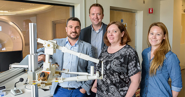
Sergi has already built an MRI-compatible, wrist-controlled robot, the MR-Softwrist, but with this grant, he’s making a new-and-improved version.
Many robots used in medical research aren’t MRI-compatible, due to their size or materials. “We are the first group in the world to have developed an MRI-compatible robot to study neural reorganization induced by robotic rehabilitation therapy,” said Sergi. In August, his team published a paper in IEEE Transactions on Biomedical Engineering that establishes a protocol to test compatibility of robots for fMRI.
Sergi is conducting this research at UD’s Center for Biomedical and Brain Imaging and at UD’s Science, Technology and Advanced Research (STAR) Campus. He will collaborate with Susanne Morton, an associate professor of physical therapy, and Curtis Johnson, an assistant professor of biomedical engineering.
Johnson looks forward to the project. “The MR-compatible robot will enable very unique experiments when used along with fMRI,” he said. “It also shows off what kind of research is possible with having the MRI scanner on campus at the CBBI.”
“This is truly multi-disciplinary research, which combines innovations in robot design and control, as well as in neuroimaging, and which we hope will have an impact in the field of clinical neuroscience,” said Sergi.
Dawn Elliott, chair of the Department of Biomedical Engineering, said: “Fabrizio’s research with MRI-compatible robots is a great example of what biomedical engineers do—create innovative technologies and apply them in ways that will improve people’s lives.”
Sergi is the director of UD’s Human Robotics lab. He received his doctoral degree in biomedical engineering from Università Campus Bio-Medico di Roma.
Contact Us
Have a UDaily story idea?
Contact us at ocm@udel.edu
Members of the press
Contact us at 302-831-NEWS or visit the Media Relations website
ADVERTISEMENT
Resources
Office of Communications & Marketing 105 E. Main St. Newark, DE 19716 ocm@udel.edu Phone: 302-831-2792