
This website contains microscopic images at both the light and electron microscopic level collected by Dr. Wagner and Hossler over their years of teaching and research in microscopic anatomy. It also contains course material in Mammalian Histology taught by Dr. Wagner. Their are also links to animations and videos pertaining to biological structure at the microscopic level.
LIGHT MICROSCOPIC IMAGES OF MAMMALIAN ORGANS AND TISSUES
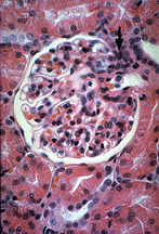
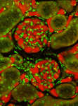
High magnification images of sections through mammalian tissue and organs. Most are stained with Hematoxylin and eosin but in several cases more specialized stains are used. |
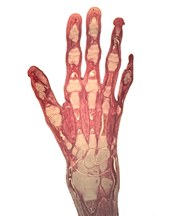
Low magnification images taken with a Leitz Photomakroscope permit surveillance of large tissue regions giving a perspective seldom achieved with a conventional compound microscope |
CELL AND TISSUE ULTRASTRUCTURE
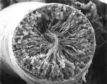 |
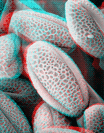
Transmission and scanning electron micrographs of mammalian tissues |
HISTOLOGY POWERPOINT PRESENTATIONS
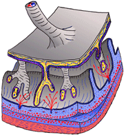
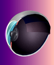
| Anatomical illustrations and power point presentations of lectures given in B408 Mammlian Histology |
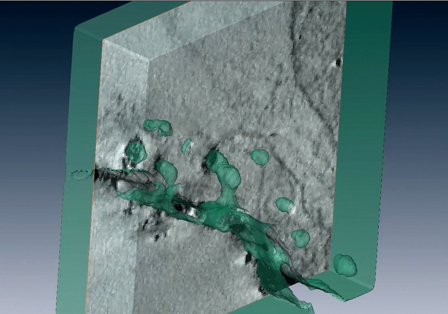
THREE DIMENSIONAL MODELS AND VIDEOS OF CELLS AND TISSUES
|
 Animated videos of models of biological tissues and structures rotated through 360 degrees to provide views from all angles Animated videos of models of biological tissues and structures rotated through 360 degrees to provide views from all angles |
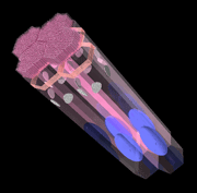
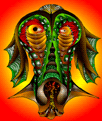
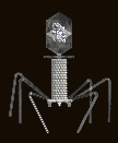
| Computer Art by Dr. Roger C. Wagner
|
 |
 |
 |
 |
 |
 |