ADVERTISEMENT
- Rozovsky wins prestigious NSF Early Career Award
- UD students meet alumni, experience 'closing bell' at NYSE
- Newark Police seek assistance in identifying suspects in robbery
- Rivlin says bipartisan budget action, stronger budget rules key to reversing debt
- Stink bugs shouldn't pose problem until late summer
- Gao to honor Placido Domingo in Washington performance
- Adopt-A-Highway project keeps Lewes road clean
- WVUD's Radiothon fundraiser runs April 1-10
- W.D. Snodgrass Symposium to honor Pulitzer winner
- New guide helps cancer patients manage symptoms
- UD in the News, March 25, 2011
- For the Record, March 25, 2011
- Public opinion expert discusses world views of U.S. in Global Agenda series
- Congressional delegation, dean laud Center for Community Research and Service program
- Center for Political Communication sets symposium on politics, entertainment
- Students work to raise funds, awareness of domestic violence
- Equestrian team wins regional championship in Western riding
- Markell, Harker stress importance of agriculture to Delaware's economy
- Carol A. Ammon MBA Case Competition winners announced
- Prof presents blood-clotting studies at Gordon Research Conference
- Sexual Assault Awareness Month events, programs announced
- Stay connected with Sea Grant, CEOE e-newsletter
- A message to UD regarding the tragedy in Japan
- More News >>
- March 31-May 14: REP stages Neil Simon's 'The Good Doctor'
- April 2: Newark plans annual 'wine and dine'
- April 5: Expert perspective on U.S. health care
- April 5: Comedian Ace Guillen to visit Scrounge
- April 6, May 4: School of Nursing sponsors research lecture series
- April 6-May 4: Confucius Institute presents Chinese Film Series on Wednesdays
- April 6: IPCC's Pachauri to discuss sustainable development in DENIN Dialogue Series
- April 7: 'WVUDstock' radiothon concert announced
- April 8: English Language Institute presents 'Arts in Translation'
- April 9: Green and Healthy Living Expo planned at The Bob
- April 9: Center for Political Communication to host Onion editor
- April 10: Alumni Easter Egg-stravaganza planned
- April 11: CDS session to focus on visual assistive technologies
- April 12: T.J. Stiles to speak at UDLA annual dinner
- April 15, 16: Annual UD push lawnmower tune-up scheduled
- April 15, 16: Master Players series presents iMusic 4, China Magpie
- April 15, 16: Delaware Symphony, UD chorus to perform Mahler work
- April 18: Former NFL Coach Bill Cowher featured in UD Speaks
- April 21-24: Sesame Street Live brings Elmo and friends to The Bob
- April 30: Save the date for Ag Day 2011 at UD
- April 30: Symposium to consider 'Frontiers at the Chemistry-Biology Interface'
- April 30-May 1: Relay for Life set at Delaware Field House
- May 4: Delaware Membrane Protein Symposium announced
- May 5: Northwestern University's Leon Keer to deliver Kerr lecture
- May 7: Women's volleyball team to host second annual Spring Fling
- Through May 3: SPPA announces speakers for 10th annual lecture series
- Through May 4: Global Agenda sees U.S. through others' eyes; World Bank president to speak
- Through May 4: 'Research on Race, Ethnicity, Culture' topic of series
- Through May 9: Black American Studies announces lecture series
- Through May 11: 'Challenges in Jewish Culture' lecture series announced
- Through May 11: Area Studies research featured in speaker series
- Through June 5: 'Andy Warhol: Behind the Camera' on view in Old College Gallery
- Through July 15: 'Bodyscapes' on view at Mechanical Hall Gallery
- More What's Happening >>
- UD calendar >>
- Middle States evaluation team on campus April 5
- Phipps named HR Liaison of the Quarter
- Senior wins iPad for participating in assessment study
- April 19: Procurement Services schedules information sessions
- UD Bookstore announces spring break hours
- HealthyU Wellness Program encourages employees to 'Step into Spring'
- April 8-29: Faculty roundtable series considers student engagement
- GRE is changing; learn more at April 15 info session
- April 30: UD Evening with Blue Rocks set for employees
- Morris Library to be open 24/7 during final exams
- More Campus FYI >>
12:11 p.m., July 20, 2010----A University of Delaware research team has received a $300,000 National Science Foundation grant to better understand ultrasound echoes of encapsulated microbubbles used for noninvasive blood pressure monitoring.
Ultrasound, which uses a pulsing high frequency sound beyond the upper limit of human hearing to peer into the body and provide images, is an important tool in modern health care.
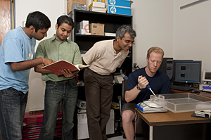
Kausik Sarkar, associate professor in the Department of Mechanical Engineering, is the principal investigator for the project.
Sarkar said microbubbles -- tiny encapsulated bubbles of gas -- are used as a contrast-enhancing agent for diagnostic ultrasound because they are small enough to penetrate deep into the human body.
The project is predicated on the use of a subharmonic signal -- a signal at a frequency lower than that of the exciting ultrasound pulse -- from these microbubbles to noninvasively quantify and monitor the ambient blood pressure in an organ.
“Local ambient blood pressure provides important information regarding the functional integrity of many organs, and it can be used to diagnose and monitor many diseases such as defective heart valves, malignant tumors and portal hypertension,” Sarkar said. “We propose to use in vitro experiments, modeling, perturbative analysis and numerical simulation to investigate the dynamics of contrast microbubbles with a view to understand and optimize such applications.”
Optimization is important because while ultrasound is the safest means of medical imaging, its utility is limited by poor contrast, Sarkar said, noting that about 20 percent of the 20 million echocardiographies performed each year in the United States do not provide definitive diagnosis for coronary heart disease.
“A good contrast agent can measurably improve ultrasound imaging,” he said, and better understanding the response of contrast microbubbles is key to that improvement.
Sarkar's research group has been working on encapsulated contrast microbubbles for a number of years and has established a collaborative research program with Thomas Jefferson University's radiology department that has secured funding from Department of Defense and the National Institutes of Health.
The UD team had a prior NSF grant and also is funded by an NIH IDeA Network of Biomedical Research Excellence (INBRE) grant.
Sarkar joined the UD faculty in 2001. He received a bachelor's degree from the Indian Institute of Technology, and a master's and doctorate from the Johns Hopkins University.
Article by Neil Thomas
Photo by Kathy F. Atkinson


