- Rozovsky wins prestigious NSF Early Career Award
- UD students meet alumni, experience 'closing bell' at NYSE
- Newark Police seek assistance in identifying suspects in robbery
- Rivlin says bipartisan budget action, stronger budget rules key to reversing debt
- Stink bugs shouldn't pose problem until late summer
- Gao to honor Placido Domingo in Washington performance
- Adopt-A-Highway project keeps Lewes road clean
- WVUD's Radiothon fundraiser runs April 1-10
- W.D. Snodgrass Symposium to honor Pulitzer winner
- New guide helps cancer patients manage symptoms
- UD in the News, March 25, 2011
- For the Record, March 25, 2011
- Public opinion expert discusses world views of U.S. in Global Agenda series
- Congressional delegation, dean laud Center for Community Research and Service program
- Center for Political Communication sets symposium on politics, entertainment
- Students work to raise funds, awareness of domestic violence
- Equestrian team wins regional championship in Western riding
- Markell, Harker stress importance of agriculture to Delaware's economy
- Carol A. Ammon MBA Case Competition winners announced
- Prof presents blood-clotting studies at Gordon Research Conference
- Sexual Assault Awareness Month events, programs announced
- Stay connected with Sea Grant, CEOE e-newsletter
- A message to UD regarding the tragedy in Japan
- More News >>
- March 31-May 14: REP stages Neil Simon's 'The Good Doctor'
- April 2: Newark plans annual 'wine and dine'
- April 5: Expert perspective on U.S. health care
- April 5: Comedian Ace Guillen to visit Scrounge
- April 6, May 4: School of Nursing sponsors research lecture series
- April 6-May 4: Confucius Institute presents Chinese Film Series on Wednesdays
- April 6: IPCC's Pachauri to discuss sustainable development in DENIN Dialogue Series
- April 7: 'WVUDstock' radiothon concert announced
- April 8: English Language Institute presents 'Arts in Translation'
- April 9: Green and Healthy Living Expo planned at The Bob
- April 9: Center for Political Communication to host Onion editor
- April 10: Alumni Easter Egg-stravaganza planned
- April 11: CDS session to focus on visual assistive technologies
- April 12: T.J. Stiles to speak at UDLA annual dinner
- April 15, 16: Annual UD push lawnmower tune-up scheduled
- April 15, 16: Master Players series presents iMusic 4, China Magpie
- April 15, 16: Delaware Symphony, UD chorus to perform Mahler work
- April 18: Former NFL Coach Bill Cowher featured in UD Speaks
- April 21-24: Sesame Street Live brings Elmo and friends to The Bob
- April 30: Save the date for Ag Day 2011 at UD
- April 30: Symposium to consider 'Frontiers at the Chemistry-Biology Interface'
- April 30-May 1: Relay for Life set at Delaware Field House
- May 4: Delaware Membrane Protein Symposium announced
- May 5: Northwestern University's Leon Keer to deliver Kerr lecture
- May 7: Women's volleyball team to host second annual Spring Fling
- Through May 3: SPPA announces speakers for 10th annual lecture series
- Through May 4: Global Agenda sees U.S. through others' eyes; World Bank president to speak
- Through May 4: 'Research on Race, Ethnicity, Culture' topic of series
- Through May 9: Black American Studies announces lecture series
- Through May 11: 'Challenges in Jewish Culture' lecture series announced
- Through May 11: Area Studies research featured in speaker series
- Through June 5: 'Andy Warhol: Behind the Camera' on view in Old College Gallery
- Through July 15: 'Bodyscapes' on view at Mechanical Hall Gallery
- More What's Happening >>
- UD calendar >>
- Middle States evaluation team on campus April 5
- Phipps named HR Liaison of the Quarter
- Senior wins iPad for participating in assessment study
- April 19: Procurement Services schedules information sessions
- UD Bookstore announces spring break hours
- HealthyU Wellness Program encourages employees to 'Step into Spring'
- April 8-29: Faculty roundtable series considers student engagement
- GRE is changing; learn more at April 15 info session
- April 30: UD Evening with Blue Rocks set for employees
- Morris Library to be open 24/7 during final exams
- More Campus FYI >>
11:08 a.m., Oct. 13, 2009----Two faculty members in the University of Delaware's Department of Electrical and Computer Engineering are part of a team that was recently awarded an $849,000 grant from the Department of Defense to develop a three-dimensional projection environment for molecular design and surgical simulation. The award was made through the U.S. Army's Telemedicine & Advanced Technology Research Center (TATRC).
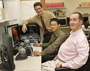
The UD team includes Kenneth Barner, professor and department chairperson; Karl Steiner, professor and associate provost for interdisciplinary research initiatives; and Rui Hu, a doctoral candidate in electrical engineering.
The lead institution on the project is Thomas Jefferson University (TJU), with the TJU team including biochemist Eric Wickstrom, who acts as principal investigator on the project, radiologist Matthew Thakur, surgeon John Kairys, medical educator Martha Ankeny, computer specialist Devakumar Devadhas, synthetic chemist Chang-Po Chen and biochemistry doctoral candidate Yuan-Yuan Jin.
The initial work will focus on the pancreas, as pancreatic cancer grows rapidly and presents difficult surgical challenges.
Under the UD component of the project, Barner and Steiner will build upon their earlier research in 3D virtual surgery simulation and work with their medical colleagues at TJU to create the next generation of a haptics-based virtual surgery simulator.
While radiologic images give surgeons a visual representation of what they may encounter at the time of exploration, current imaging systems do not provide genetic information or tactile information about the tissues that will be encountered at surgery, nor do they allow physical interaction with the image.
“Haptics provides tactile, or touch, feedback to the user via a small robot that is integrated with the visual simulation on the screen,” Steiner explains. “As the user moves the robot, a simulated object, such as a scalpel or other surgical instrument, moves within the 3-D environment, which includes simulations of various organs in the human body.”
The organ simulations have been generated through a process called segmentation, where data taken from anatomical CT scans and molecular or genetic PET scans are digitally processed slice-by-slice to extract the outlines of individual organs in a patient.
Once the internal structure of the body has been segmented, that data is processed and the organs are integrated with a volumetric simulation that depicts mechanical properties, for example, healthy tissue or diseased tissue in a lung or pancreas. Next, the biochemical activity inside cancer cells is fused with the anatomical image.
The haptics interface then allows manipulation of the surgical instruments, and, as the instrument touches one of the simulated organs, the deformation of the organ is calculated and visualized. Information is provided to the haptics robot as to the forces required to push on the organ. Tumor texture will be represented as firmer than normal, healthy pancreas.
“This environment is the basis for our new collaboration with TJU,” says Steiner, “where we will now focus on a set of data from CT and PET scans provided by TJU. We will merge these datasets to provide a scene with state-of-the-art information about the disease state of the organ.”
The project is yet another link among research groups at UD and TJU, and the research benefits from and contributes to the growing collaboration under the Delaware Health Sciences Alliance.
“The unique aspect of this project,” Barner says, “is that it enables us to build on our prior results for deformable objects in surgery simulation by partnering with researchers at Jefferson. Our aim is not only to take surgery simulation to the next level, including the realistic interaction of multiple surgical tools and organs, but also to incorporate information from multiple imaging modalities to provide doctors with a comprehensive environment from which surgeries can be practiced and planned.”
“The project is also a great opportunity for our graduate students,” he adds, “as engineers will find it increasingly important to work with professionals from other fields as technology becomes more complex and as its applications broaden.”
Article by Diane Kukich
Photo by Ambre Alexander


