- Rozovsky wins prestigious NSF Early Career Award
- UD students meet alumni, experience 'closing bell' at NYSE
- Newark Police seek assistance in identifying suspects in robbery
- Rivlin says bipartisan budget action, stronger budget rules key to reversing debt
- Stink bugs shouldn't pose problem until late summer
- Gao to honor Placido Domingo in Washington performance
- Adopt-A-Highway project keeps Lewes road clean
- WVUD's Radiothon fundraiser runs April 1-10
- W.D. Snodgrass Symposium to honor Pulitzer winner
- New guide helps cancer patients manage symptoms
- UD in the News, March 25, 2011
- For the Record, March 25, 2011
- Public opinion expert discusses world views of U.S. in Global Agenda series
- Congressional delegation, dean laud Center for Community Research and Service program
- Center for Political Communication sets symposium on politics, entertainment
- Students work to raise funds, awareness of domestic violence
- Equestrian team wins regional championship in Western riding
- Markell, Harker stress importance of agriculture to Delaware's economy
- Carol A. Ammon MBA Case Competition winners announced
- Prof presents blood-clotting studies at Gordon Research Conference
- Sexual Assault Awareness Month events, programs announced
- Stay connected with Sea Grant, CEOE e-newsletter
- A message to UD regarding the tragedy in Japan
- More News >>
- March 31-May 14: REP stages Neil Simon's 'The Good Doctor'
- April 2: Newark plans annual 'wine and dine'
- April 5: Expert perspective on U.S. health care
- April 5: Comedian Ace Guillen to visit Scrounge
- April 6, May 4: School of Nursing sponsors research lecture series
- April 6-May 4: Confucius Institute presents Chinese Film Series on Wednesdays
- April 6: IPCC's Pachauri to discuss sustainable development in DENIN Dialogue Series
- April 7: 'WVUDstock' radiothon concert announced
- April 8: English Language Institute presents 'Arts in Translation'
- April 9: Green and Healthy Living Expo planned at The Bob
- April 9: Center for Political Communication to host Onion editor
- April 10: Alumni Easter Egg-stravaganza planned
- April 11: CDS session to focus on visual assistive technologies
- April 12: T.J. Stiles to speak at UDLA annual dinner
- April 15, 16: Annual UD push lawnmower tune-up scheduled
- April 15, 16: Master Players series presents iMusic 4, China Magpie
- April 15, 16: Delaware Symphony, UD chorus to perform Mahler work
- April 18: Former NFL Coach Bill Cowher featured in UD Speaks
- April 21-24: Sesame Street Live brings Elmo and friends to The Bob
- April 30: Save the date for Ag Day 2011 at UD
- April 30: Symposium to consider 'Frontiers at the Chemistry-Biology Interface'
- April 30-May 1: Relay for Life set at Delaware Field House
- May 4: Delaware Membrane Protein Symposium announced
- May 5: Northwestern University's Leon Keer to deliver Kerr lecture
- May 7: Women's volleyball team to host second annual Spring Fling
- Through May 3: SPPA announces speakers for 10th annual lecture series
- Through May 4: Global Agenda sees U.S. through others' eyes; World Bank president to speak
- Through May 4: 'Research on Race, Ethnicity, Culture' topic of series
- Through May 9: Black American Studies announces lecture series
- Through May 11: 'Challenges in Jewish Culture' lecture series announced
- Through May 11: Area Studies research featured in speaker series
- Through June 5: 'Andy Warhol: Behind the Camera' on view in Old College Gallery
- Through July 15: 'Bodyscapes' on view at Mechanical Hall Gallery
- More What's Happening >>
- UD calendar >>
- Middle States evaluation team on campus April 5
- Phipps named HR Liaison of the Quarter
- Senior wins iPad for participating in assessment study
- April 19: Procurement Services schedules information sessions
- UD Bookstore announces spring break hours
- HealthyU Wellness Program encourages employees to 'Step into Spring'
- April 8-29: Faculty roundtable series considers student engagement
- GRE is changing; learn more at April 15 info session
- April 30: UD Evening with Blue Rocks set for employees
- Morris Library to be open 24/7 during final exams
- More Campus FYI >>
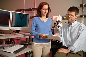
8:26 a.m., Jan. 7, 2009----Kirk Czymmek, associate professor of biological sciences at the University of Delaware, and associate scientist Elizabeth Adams, researchers in the Bio-Imaging Center at the Delaware Biotechnology Institute, have won a grant for a new technology add-on to the facility's BioScope II.
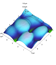
The BioScope is a Scanning Probe Microscope (SPM) that creates three-dimensional pictures of surfaces, allowing scientists to examine topography, phase, and force interactions in samples.
“The microscope add-on is literally an electronic module, with incredibly powerful capabilities,” said Adams, who also has a secondary appointment as assistant professor in the University of Delaware's Department of Biological Sciences.
This new component, Adams explained, serves a highly specific purpose -- in technical terms, it measures elasticity. “If you change something about your sample, does elasticity change?”
One factor in elasticity is determined by the strength of neighboring molecules and their precise arrangement on any surface, whether natural or synthetic. “To appreciate this in real-world terms, it is like the difference in the sensation you get when walking on the grass versus pavement, but at an atomic level,” said Adams.
“The acquisition of this new technology is a major accomplishment for the University of Delaware and its partners,” added Czymmek, director of the Bio-Imaging Center. “The new capability will extend to a broad array of materials and biological applications. What I am particularly excited about is its ability to map molecular interactions of cell surfaces over 1,000 times faster than current technologies. We plan to work very closely with the developers, Veeco Labs, to expand the application of this technology into new frontiers of biological analysis.”
Officially called the HarmoniX Nanoscale Material Mapping upgrade module, the enhancement is the first atomic force microscope (AFM) to map, on the nanoscale, different materials' properties, such as adhesion, stiffness and average force.
HarmoniX is unique because its property maps are both real-time and high-resolution. It allows scientists to characterize soft materials, thin films, small particles, or domains within a bulk solid.
Veeco Labs, creators of HarmoniX, established the HarmoniX Innovation Research Grant Program in 2008, which provides winners with the hardware, software, training and installation of the HarmoniX module, a $25,000 value.
Scientists who run scanning probe microscopes were encouraged to submit their ideas for 12-month research projects using the module. Adams said that many of the proposals were in the field of materials development; UD's submission was one of the few with life science applications.
Czymmek and Adams plan to investigate fungal cell walls, which have become attractive targets for antifungal chemotherapy. They will use their new module to investigate the effects of antifungal drugs on fungal cell wall elasticity.
The researchers said they expect the data will make an important contribution to the plant and medical fields as a tool for evaluating the effects of some anti-fungal drugs.
The Bio-Imaging Center is a multi-user microscopy facility with state-of-the-art electron, confocal and light microscopes.
Article by Katie Ginder-Vogel


