CHEM-342 - INTRODUCTION TO BIOCHEMISTRY
FINAL EXAMINATION - PART I (Individual Work)
Monday, 24 May 1999, 7:00 - 9:00 p. m., 208 Gore Hall
H. B. White - Instructor
Important - Please read this before you turn the page.
B. (15 points) Short Summary Statements
C. (10 points) Problem to Solve
D. (8 points) Essay Question about the seminar you attended
E. (12 points) Application of principles to a new situation
F. (15 points) Essay
Part II, the group part of the examination, has one problem worth 25 points.
_______________________ 1. Color of a solution of oxyhemoglobin.
_______________________ 2. Color of a solution of oxyhemoglobin after it has reacted with dithionite, S2O4=.
_______________________ 3. Color of a solution of methemoglobin.
_______________________ 4. Color of a solution of methemoglobin after it has reacted with dithionite, S2O4=.
_______________________ 5. Ionizable functional group associated with histidine.
_______________________ 6. Addictive alkaloid that can be chemically converted into the vitamin niacin.
_______________________ 7. Bilirubin is associated with this symptom of sickle cell anemia as well as with hepatitis.
_______________________ 8. Sulfur-containing amino acid in proteins.
_______________________ 9. Number of heme groups in Hb S.
_______________________ 10. South American country closest to Grenada.
_______________________ 11. Bond hydrolyzed by trypsin.
_______________________ 12. Principle on which Longsworth Scanning is based.
_______________________ 13. The guanido groups is associated with this amino acid.
_______________________ 14. Functional group in proteins that reacts with Sanger's Reagent.
_______________________ 15. In the one-letter abbreviations,
"E" refers to this amino acid.
1. Herrick (1910)
2. Diggs et al. (1933)
3. Pauling et al. (1949)
4. Ingram (1959)
5. Allison (1954)
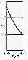
C. Problem (10 points) Consider the figure below from Svedberg and Fåhraeus (1926) which shows the concentration of hemoglobin as a function of the distance from the center of rotation in a centrifuge. What would the profile look like if hemoglobin had a molecular weight of 16,700 rather than 66,800 and the rotational velocity were doubled? Draw a line on the figure representing your conclusion and justify it in words.
From the information provided, predict the number of bands and the
relative amounts of each band in an electrophoretic separation of LDH
from a tissue producing equal amounts of H and L subunits. Assume the subunits
randomly associate to form active, non-dissociating tetramers. Describe
your reasoning and then draw a figure depicting the appearance of
an electrophoretic separation of LDH from this hypothetical tissue.Predict
the relative amounts of LDH in each band?
As you read the two abstracts of recent articles on the following page,
keep track of the things you don't understand and the thoughts you have.
(Use the margins for notes, if you want.) Collect
your thoughts and write in the space below a one-page narrative that integrates
the new information with things you know and the things you would like
to
know relating to the topic. I am looking for a well organized and well
articulated presentation of your personal reflections rather than a summary
or paraphrasing of information in the abstracts. You may continue on the
back of this page, if you need more space.
Blood 85(4): 1111-1117 (1995)
An analysis of fetal hemoglobin variation in sickle cell disease: the relative contributions of the X-linked factor, beta globin haplotypes, alpha-globin gene number, gender, and age.
Chang YC, Smith K, Moore RD, Serjeant GR and Dover GJ.
Department of Pediatrics, Johns Hopkins University School of Medicine, Baltimore, MD.
Five factors have been shown to influence the 20-fold variation of fetal hemoglobin (Hb F) levels in sickle cell anemia (SS): age, sex, the alpha-globin gene number, beta-globin haplotypes, and an X-linked locus that regulates the production of Hb F-containing erythrocytes (F cells), ie, the F-cell production (FCP) locus. To determine the relative importance of these factors, we studied 257 Jamaican SS subjects from a Cohort group identified by newborn screening and from a Sib Pair study. Linear regression analysis showed that each variable, when analyzed alone, had a significant association with Hb F levels (P<.05). Multiple regression analysis, including all variables, showed that the FCP locus is he strongest predictor, accounting for 40% of Hb F variation. Beta-globin haplotypes, alpha-globin genes, and age account for less han 10% of Hb F variation. The association between the beta-globin haplotypes and Hb F levels becomes apparent if the influence of the FCP locus s removed by analyzing only individuals with the same FCP phenotype. Thus, the FCP locus is the most important factor identified to date in determining Hb F levels. The variation within each FCP phenotype is modulated by factors associated with the three common beta-globin haplotypes and other yet identified factor(s)
Blood 93(6): 1790-1797 (1999)
Sustained induction of fetal hemoglobin by pulse butyrate therapy in sickle cell disease
Atweh GF, Sutton M, Nassif I, Boosalis V, Dover GJ, Wallenstein S, Wright E, McMahon L, Stamatoyannopoulos G, Faller DV and Perrine SP.
Departments of Medicine, Pediatrics and Biomathematical Sciences, Mount Sinai School of Medicine, New York, NY
High levels of fetal hemoglobin (Hb F)
protect from many of the complications of sickle cell disease and lead
to improved survival. Butyrate and other short chain fatty acids were previously
shown to increase Hb F production in erythroid cells in vitro and in animal
models in vivo. However, butyrates are also known to inhibit the proliferation
of many cell types, including erythroid cells. Experience with the use
of butyrate in animal models and in early clinical trials demonstrated
that the Hb F response may be lost after prolonged administration of high
doses of butyrate. We designed a regimen consisting of intermittent or
pulse therapy in which butyrate was administered for 4 days followed by
10 to 24 days with no drug exposure. This pulse regimen induced fetal hemoglobin
expression in 9 of 11 patients. The mean Hb F in this group increased from
7.2% to 21.0% (P<.002) after intermittent butyrate therapy for a mean
duration of 29.9 weeks. This was associated with a parallel increase in
the number of F cells and F reticulocytes. The total hemoglobin levels
also increased from a mean of 7.8 g/dL to a mean of 8.8 g/dL (P<.006).
The increased levels of Hb F were sustained in all responders, including
1 patient who has been on butyrate therapy for more than 28 months. This
regimen, which resulted in a marked and sustained increase in Hb F levels
in more than two thirds of the adult sickle cell patients enrolled in this
study, was well tolerated without adverse side effects. These encouraging
results require confirmation along with appropriate evaluation of clinical
outcomes in a larger number of patients with sickle cell disease.
FINAL EXAMINATION - PART II (Group Work)
Monday 24 May 1999, 9:00 - 10:00 PM
H. B. White - Instructor
(25 points total) The abstract below accompanied an article published six months ago in the British Journal of Hematology. Read it carefully and then answer the questions that follow.
Br J Haematol 103(4): 950-956 (1998)
A new sickle cell disease phenotype associating Hb S trait, severe pyruvate kinase deficiency (PK Conakry), and an alpha2 globin gene variant (Hb Conakry).
Cohen-Solal M, Prehu C,
Wajcman H, Poyart C, Bardakdjian-Michau J, Kister J, Prome D, Valentin
C, Bachir D, and Galacteros F. INSERM U474, Hopital Henri Mondor, Creteil,
France
Guinean woman, heterozygous for hemoglobin
(Hb) S, was studied because of episodes of marked anaemia, repeated typical
metaphyseal painful crises and hemosiderosis. Her sickling syndrome resulted
from the association of Hb S trait with severe pyruvate kinase deficiency
leading to a 2,3-DPG concentration twice normal levels. Sequence of the
PK-R gene revealed an undescribed mutation in the homozygous or hemizygous
state within exon 5 (nucleotide 2670 C A), leading to the interchange of
Ser 130 into Tyr (PK Conakry). In addition, the patient carried a new hemoglobin
variant, Hb Conakry [alpha80(F1) Leu Val], which seemed to have a mild
effect. The high intraerythrocytic 2,3-DPG concentration induced by the
PK deficiency resulted in a decreased oxygen affinity which favored sickling
to a level almost similar to that of Hb S/C compound heterozygous patients.
This was confirmed by oxygen binding measurements of Hb A/Hb S erythrocytes
in which 2,3-DPG content was modified in vitro. Hysteresis between deoxy-
and reoxygenation curves, as well as increase in the n(max) value, demonstrated
that the extent of Hb S polymerization in the propositus was almost the
same as that of RBCs from a homozygous sickle cell patent or those of an
A/S heterozygous patient with an artificial increase of 2,3-DPG concentration.
A. (10 points) Predict the appearance
of the Hb electrophoretic pattern from the patient compared to normal Hb
A and people with classical sickle cell anemia Hb S. Draw a picture and
explain your reasoning.
B. (15 points) Based on the information provided in the abstract, draw oxygen-binding curves (% Hb saturation vs pO2) for erythrocytes from individuals with Hb S, Hb A/S, Hb S/C, and the patient. Indicate the normal range of pO2 in the body and, where relevant, note where sickling begins to occur. Explain your reasoning. Conceptual understanding and relationships here are more important than specific numbers.