|
|
 EB2014 EXPERIMENTAL BIOLOGY MEETINGS San Diego, CA APRIL 26-30, 2014  |
 |
|
|
EB2014 EXPERIMENTAL BIOLOGY MEETINGS San Diego, CA APRIL 26-30, 2014 |
|
The University of Delaware group included four faculty and 9 undergraduates.
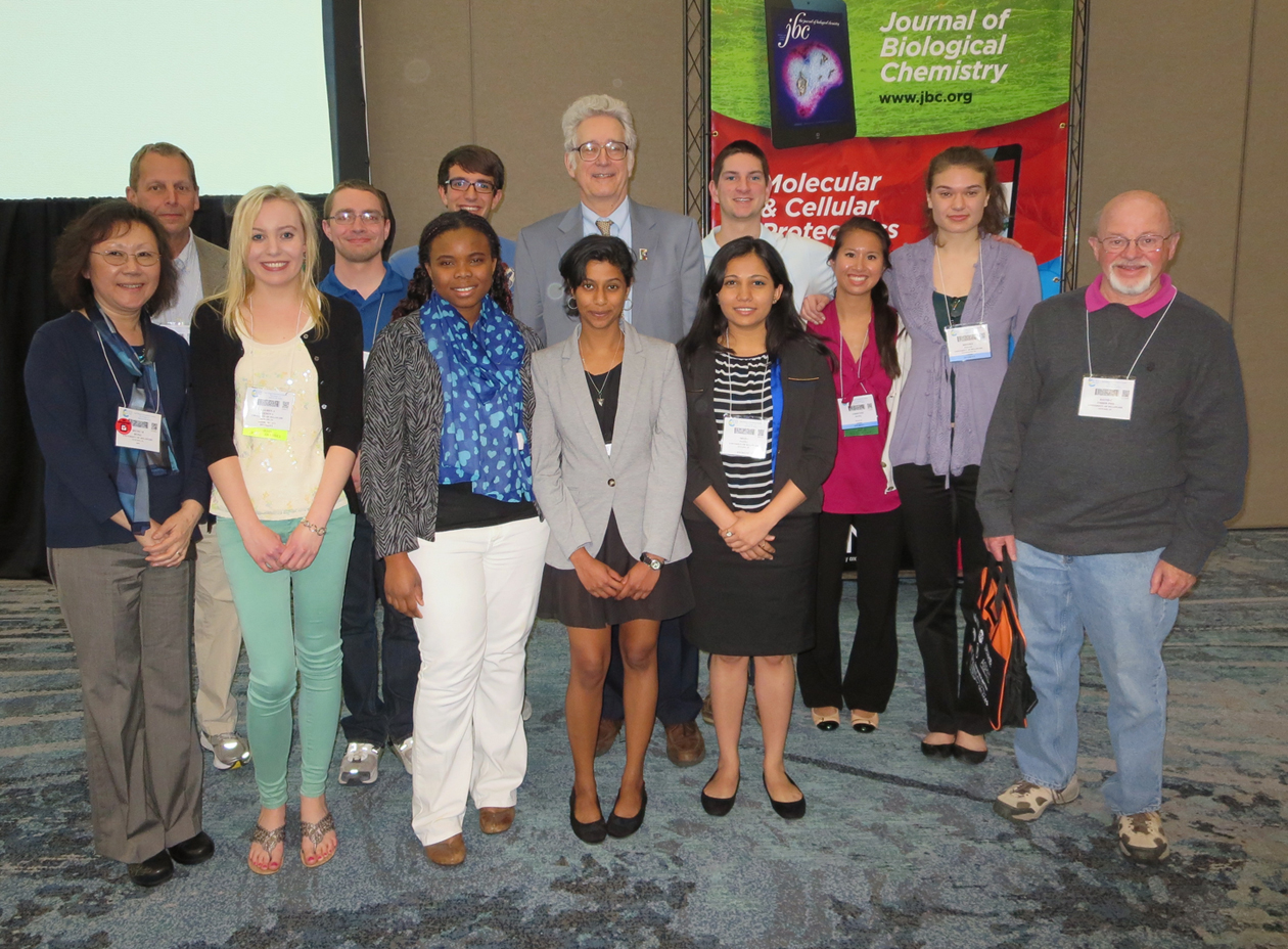 Back row, left to right: Gary Laverty, Seth Ritter, Doug Kenny, Hal White, Matt Urbam, Christine Dang, and Brooke Palus. |
||
| Prof.
Hal White, Chem & Biochem Prof. Dave Usher, Biol. Sci. Prof. Seung Hong, Biol Sci Prof. Gary Laverty, Biol Sci |
Christine
Dang, Biological Sciences Lauren Genova, Chemistry Fanta Kalle, Biological Sciences Douglas Kenny, Chemistry Brooke Palus, Biological Sciences |
Shaili
Patel, Biological Sciences Seth Ritter, Chemical Engineering Ramya Sridharan, Biochemistry Matthew Urban, Biochemistry |
|
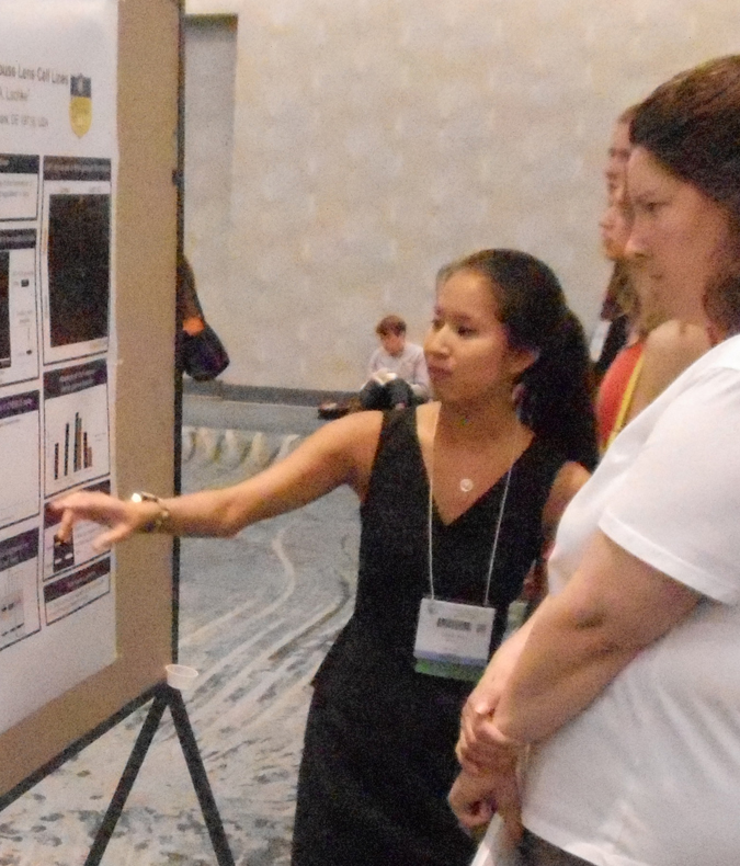
Characterization of Celf1 function in
mouse lens cell lines Although the human genome encodes ~1500 RNA binding protein (RBP) encoding genes, mutations in only ~20 are associated with human disease. We used a novel bioinformatics approach iSyTE (integrated Systems Tool for Eye gene discovery) to identify a new RBP, Celf1 (CUGBP Elav (embryonic lethal, abnormal vision)-Like Family member 1), in the lens that is potentially associated with cataract. Celf1 contains three conserved RNA Regulatory Motifs that function in sequence-specific binding to target RNAs. To develop a new resource to study Celf1 function in lens cells, we generated and characterized Celf1-knockdown (KD) mouse lens epithelial cell lines. We first selected the mouse lens epithelial cell line 21EM15 as it expresses several critical lens-expressed genes and thus recapitulates important aspects of lens biology. We infected 21EM15 cells with lentiviral particles carrying inhibitory short hairpin (sh)RNAs against mouse Celf1 mRNA and isolated infected cells based on their acquired resistance to puromycin. Celf1-KD was confirmed by reduced Celf1 expression in Western blotting. We are presently characterizing the effect of Celf1-KD on distinct classes of RNA granules (e.g. Stress granules and Processing bodies) involved in post-transcriptional regulation. In sum, we describe development of a new Celf1-KD lens cell line as a unique resource for gaining novel functional insights of an RBP in lens biology. Poster: Mon, Apr 28, 1:15 - 2:45 PM 746.1/D62 - |
|
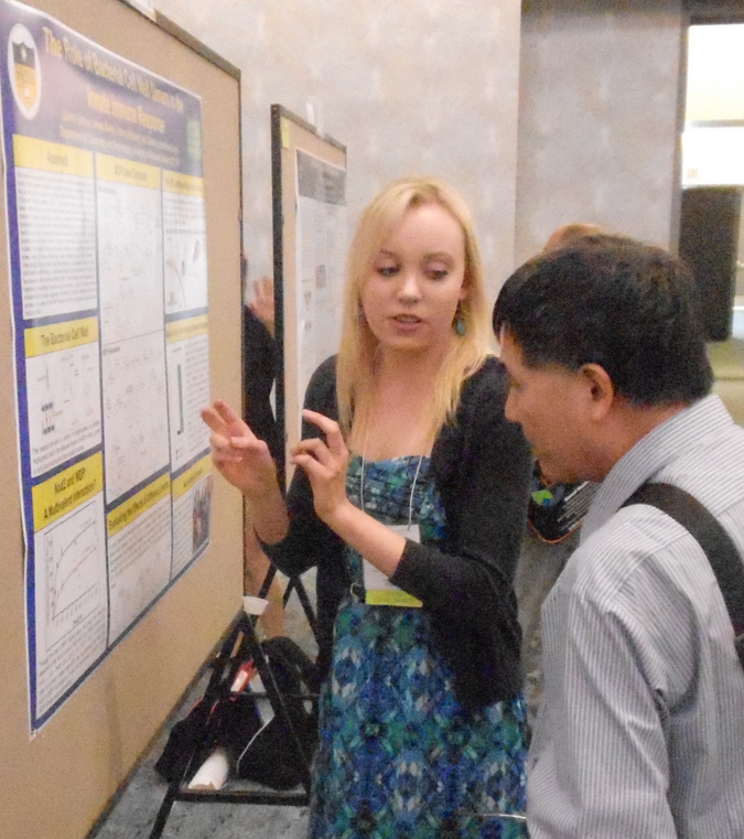
The Role of Bacterial Cell Wall Dimers in
the Innate Immune Response
Department of Chemistry and Biochemistry, University of Delaware, Newark, DE 19716 The innate immune
system is the body’s first line of defense against pathogens. The
innate immune system is triggered by pathogen associated molecular
patterns (PAMPs) that are recognized by pattern recognition receptors
(PRRs) such as Toll-like receptors (TLRs) and Nod-like receptors
(NLRs). This research project focuses on providing a better
understanding of how the innate immune system senses and responds to
the presence of bacteria. Specifically, our group is interested in the
relationship between Nucleotide-binding oligomerization
domain-containing protein 2 (Nod2), an NLR protein found in the cytosol
of mammalian host cells, and muramyl dipeptide (MDP), the smallest
bacterial cell wall fragment known to elicit an immunological response.
When Nod2 is mutated, the signaling pathway becomes disrupted and
uncontrollable inflammation arises, leading to chronic inflammatory
bowel disorders such as Crohn’s disease. In order to discover how to
better treat these diseases, it is imperative to learn more about
how Nod2 and MDP interact, a mechanism which is currently unknown. The
Grimes Lab has previously shown that Nod2 binds to MDP in vitro;
however, research suggests that a heightened immunological response may
be elicited in a host if molecules containing multiple MDP’s are used,
suggesting multivalency is at play. This project highlights my current
research progress to synthesize a variety of novel MDP dimers to test
for a multivalent interaction between Nod2 and MDP.
|
|
|
Effects of heparin and heparin-binding growth factor on human aortic adventitial fibroblasts
1Biological Sciences,
University of
Delaware, Newark, DE; 2Biomedical
Research, Nemours - Alfred I. duPont Hospital for Children, Wilmington,
DE; 3Materials Science & Engineering, University of
Delaware, Newark, DE
Poster: Tue, Apr 29, 1:15 - 2:45 PM 1012.4/D376 Recipient of an ASBMB-UAN $500 Undergraduate Travel Award.
|
|
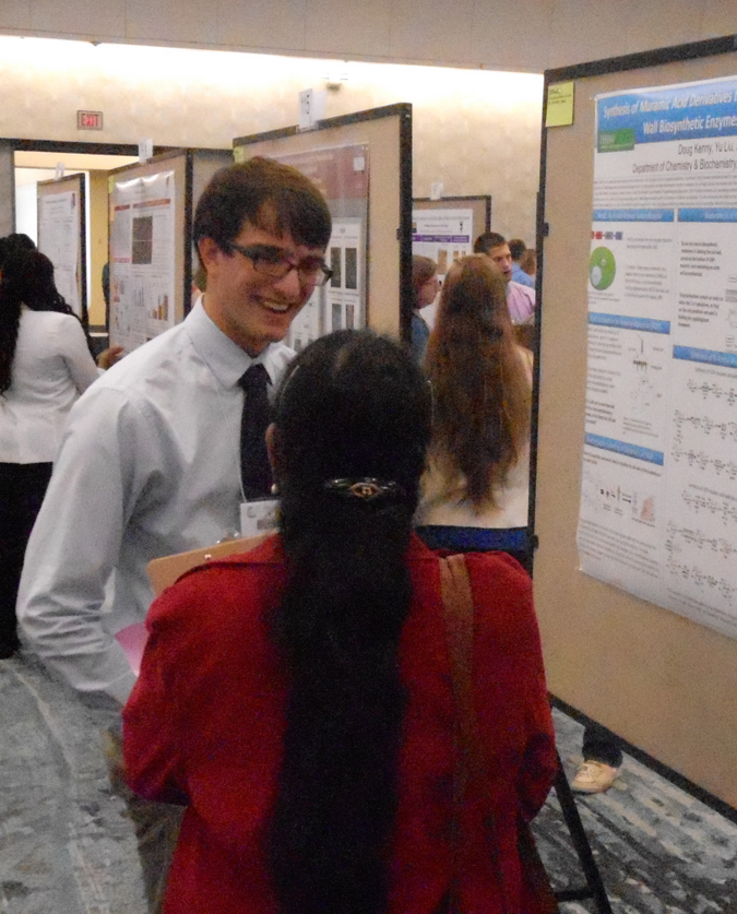
Synthesis of Muramic Acid
Derivatives to Investigate the Promiscuity of Bacterial Cell Wall
Biosynthetic Enzymes for the Investigation of Nod2
Douglas Kenny, Yu Liu, Catherine
L. Grimes
Department of Chemistry and Biochemistry, University of Delaware Poster: Tue, Apr 29, 12:15 - 1:45 PM 970.2/D179 |
|
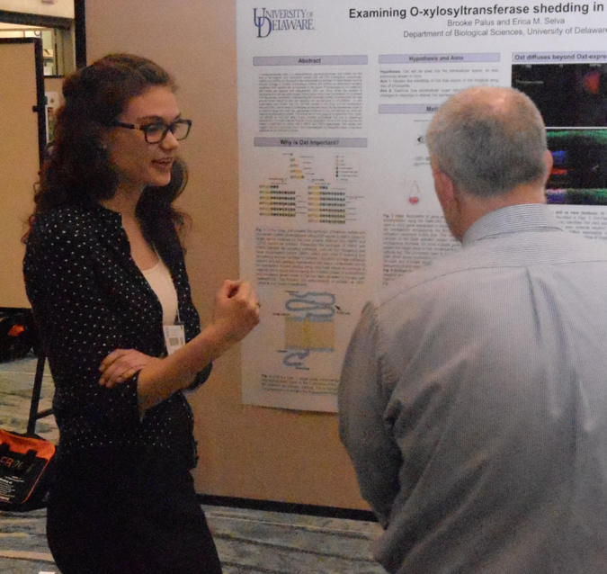
Examining
O-xylosyltransferase shedding in Drosophila
Brooke Palus and Erica Selva Department of Biological Scences, University of Delaware, Newark, DE O-xylosyltransferase (Oxt) is a Golgi transmembrane glycosyltransferase that initiates the first step in the heperan and chrondroitin sulfate biosynthesis on core proteoglycans (HSPG and CSPG). HSPGs and CSPGs are abundant extracellular matrix proteins present in virtually all tissues and have critical functions in articular cartilage in vertebrates. In this tissue, HSPGs and CSPGs are essential in regulating signaling and homeostasis. Disrupted homeostasis of articular cartilage leads to its degradation and osteoarthritis (OA). The onset of OA has been shown to result in increased serum levels of Oxt in mouse and humans. While the biosynthetic activity of Oxt has been studied, little is known about its function extracellularly. Understanding how and why Oxt is shed and its role extracellularly may provide further insight into how cell signaling and tissue homeostasis can be disrupted in OA patients. The goal of this study is to determine if Oxt is shed from Drosophila wing imaginal discs and what function extracellular Oxt has in organismal development. As expected, Oxt localizes to the Golgi and the ER the site of HSPG and CSPG biosynthesis. Oxt was also found extrcellularly suggesting it is shed in Drosophila. In loss-of-function and over expression studies the composition of the extracellular matrix and Oxt diffusion from it cell of origin were also examined. Poster: Sun, Apr 27, 12:15 - 1:45 PM 539.1/B62 |
|
Identification
of differentially expressed transcripts in Tdrd7 null mutant mouse lens
Shaili D. Patel, Carrie Barnum, Salil A. Lachke Department of Biological Sciences, Universirty of Delaware Lens is a transparent tissue within the eye that focuses light on the retina and facilitates high-resolution vision. Cataract is an eye disease that results due to the loss of lens transparency and causes severely impaired vision. It can be caused by genetic changes, aging or physiological conditions like diabetes. Treatment depends upon the patient's specific visual needs and often involves cataract surgery. Over 77 million individuals are affected worldwide, and in the United States alone, costs exceed $3 billion annually. Recently, a mutations in the human TDRD7 (Tudor domain containing protein 7) gene were shown to cause congenital/pediatric cataracts in patients. Moreover, it was also demonstrated that Tdrd7 targeted germline knockout or ENU-induced knockout mouse mutants exhibit cataracts that closely resemble the human phenotype. My research goal is to understand the regulatory function of Tdrd7 in the lens. Specifically, I aim to investigate if Tdrd7 deficiency affects the mRNA profiles in the apical and the basal regions of differentiating lens fiber cells. To achieve this I have expanded the Tdrd7 germline knockout mouse mutant colony and collected Tdrd7 mutant and control (Tdrd7+/- mice that do not exhibit cataracts) embryonic lens tissue for analysis. To study the expression of mRNAs in different locations within fiber cells, I have undertaken standardization of an approach using Laser Capture Microdissection (LCM). LCM on embryonic lens sections from Tdrd7 null mutants and control will be used to isolate the apical and the basal fiber cell regions at embryonic stage E10.5, E11.5, and E12.5. Once the specific tissues are collected, expression of mRNAs will be tested by whole genome expression profiling using Gene expression analysis. Comparative analysis of gene expression profiles of the apical and basal tissues from mutant and control will allow the identification of transcripts that are misregulated as a result of Tdrd7 mutation. Poster: Mon, Apr 28, 1:15 - 2:45 PM 747.1/D64 Recipient of a University of Delaware Unbdergraduate Research Travel Award. |
|
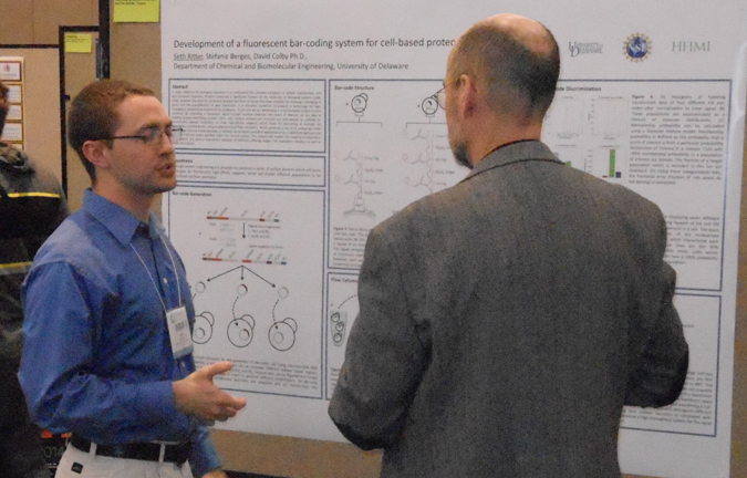
Development
of a fluorescent bar-coding system for cell-based proteomic libraries
Seth C. Ritter, Stefanie Berges, David Colby University of Delaware, Newark, DE A major objective for biological scientists is to understand the complex intricacies of cellular maintenance, and environmental response. Proteins constitute a significant fraction of the molecules in biological systems under study; however the tools for proteomic analysis fall short of those that have enabled the thorough cataloging of genes and the quantification of gene transcripts. It is therefore beneficial to produce a technology for high-throughput, quantitative analysis of the proteome. To this aim we are developing a novel heritable surface protein construct. Such surface proteins will protrude out and operate as scaffolds for fluorescently labeled antibodies, or other high-affinity reagents. By varying the number of repeating epitope elements in the scaffold a series of fluorescent signals through multiple channels and levels of intensity are generated. These Unique Identifying Proteins (UIP), allow for identification of multiple cell populations simultaneously using multidimensional flow cytometry. We found that eight unique barcodes were possible utilizing three total epitopes, where in any channel at least 60% of cells of a population could be taken while error rates in population identification remained below 5%. This system will enable the coupling of population identification and various fluorescent readouts of interests, offering insight into population statistics as well as single cell analysis. Poster: Mon, Apr 28, 12:45 - 3:00 PM 912.13/A616 |
|
Identifying
cytokines associated with acute and chronic Lyme arthritis
Ramya Sridharan1, Victoria L. Maduskie2, Paul T. Fawcett2 1Biology, University of Delaware, Hockessin, DE, 2Immunology Laboratory, Nemours/Alfred I. DuPont Hospital for Children, Wilmington, DE Symptoms of untreated infection with Borrelia burgdorferi, which is the causative agent of Lyme disease, frequently include arthritis, particularly of large joints. In pediatric patients, symptoms of arthritis typically resolve following treatment with an adequate course of antibiotics. However, in 10 to 20 percent of pediatric patients with Lyme arthritis, symptoms persist despite patients having received adequate treatment with antibiotics. Patients whose symptoms resolve in less than 6 months after initiation of antibiotic therapy are classified as having acute Lyme arthritis. Patients whose symptoms do not resolve within 6 months are classified as having chronic Lyme arthritis. Cells were obtained from the synovial fluid from pediatric patients with both chronic and acute Lyme arthritis. These were analyzed to determine if cytokines produced by synoviocytes could be used to predict the outcome of treatment with antibiotics. Primary cell cultures were cultured in transformation media to change fibroblasts into adipocytes and osteocytes. Culture supernatants were then tested for cytokine concentration changes. Results showed that transformation of synoviocytes caused significant variation of inflammatory cytokine concentrations associated with arthritis inflammation: MCP-1 and IL-6. IL-6 rapidly declined in both adipogenic and osteogenic transformations. MCP-1 also declined. Results confirm that transformation of synoviocytes from synovial fluid samples caused significant variation of inflammatory cytokine concentrations associated with arthritis inflammation. |
|
Novel
ubiquitin binding sites of S.
cerevisiae polymerase η Matthew Urban, Kun Yang, Zhihao Zhuang Dept.
of Chemistry and Biochemistry, University of Delaware, Newark, DE 19716
DNA translesion
synthesis is carried out by specialized DNA polymerases, called
Y-family
polymerases, such as Polη.
Polη contains a ubiquitin-binding zinc finger (UBZ) domain required for
effective recruitment to the stalled replication fork in response to
DNA
damages. Previous study reported a novel ubiquitin-binding mode of S.
cerevisiae
Polη in which the
C-terminal portion of Polη binds ubiquitin with 30-fold higher binding
affinity
than the UBZ domain. However, the mechanism of such higher binding
affinity is
not clear. In this study, a genetically encoded photoreactive probe, p-benzoyl-L-phenylalanine
(pBpa), was incorporated at
various positions on ubiquitin to map the interaction sites between
Polη and
ubiquitin. Data suggests that besides the I44-V70 hydrophobic patch
which was
known to bind UBZ domain of Polη, several other residues at both
C-terminus
(L73 and R74) and the back surface (I13, T14 and D24) of ubiquitin also
bind to
Polη in the C-terminus region outside the UBZ domain. These new binding
sites increase the
binding affinity between
ubiquitin and Polη, which could be
important for understanding of the role of ubiquitin in the recruitment
of Polη
during DNA transelsion synthesis. Poster:
Tue, Apr 29, 1:15 - 2:45 PM 927.4/D14 |
|
Stimulating
Attitudes of Inquiry
Harold B. White Department of Chemistry and Biochemistry, Univ. of Delaware, Newark, DE Much of contemporary science education focuses on what we know, but neglects the evidence supporting the conclusions that fill textbooks. Students thus often learn information as abstractions disconnected from their experience. They rarely encounter the observations, clever experiments, and controversies that generate new ideas and refute old. Problem-based learning (PBL) is an approach to education that encourages students to identify what they don’t know, ask questions, and actively pursue knowledge based on their curiosity and through collaboration with other students. Rather than attempt to cover massive amounts of information, PBL seeks to foster life-long habits of learning through in-depth exploration and fundamental understanding of carefully selected problems that reveal basic concepts and interdisciplinary connections. Problems based on the primary literature with emphasis on classic articles expose students not only to the logic of research but introduce them to the history of science, the people who made important discoveries, and the culture science. Recipient of ASBMB Award for Excemplary Contributions to Education -see pg 27 Award Lecture: Sun, Apr 27, 12:50 - 1:20 PM 100.1 Video recording of award lecture |
 No wonder we couldn't find your poster. |
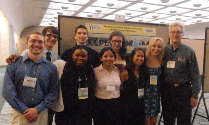 UD Group in front of UD ASBMB-Undergraduate Affiliate Poster. |
 Plenary Speakers for ASBMB |
|
 Doug marking time before takeoff at PHL. |
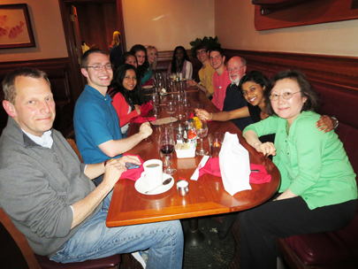 Dinner at the Fish market in San Diego. |
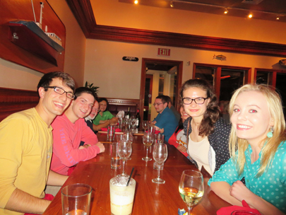 Dinner at the Fish market in San Diego. |
 Checking cell phones upon arrival in San Diego. |
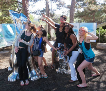
 Defiying gravity in San Diego. 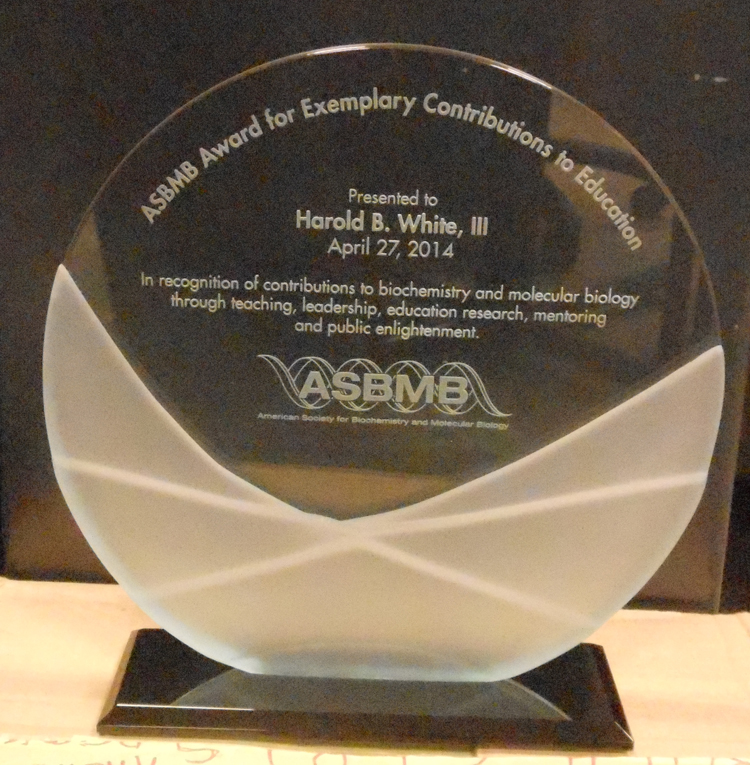 Dr. White's Award |
 Finally, Palms trees again after 10 years! |
 The tatoos are only temporary. |
 Experimental Biol;ogy 2014 |
 Selfie. What's Doug contemplating? |
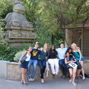  At the Zoo |
 Seth and Ramya on Coranado Island across from the Conference Center. |
|
 With a pool like this, who wants to go to conference session? |
 Pretty nice view from Hotel room. |
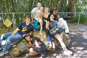 This hippo isn't very soft |
|
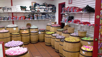 Candy Store! |
 Ticket to Old Town |
 Lauren and Fanta |
|
The trip to the Experimental Biology
2014 Meetings
in San Diego was organized by the University of Delaware HHMI
Undergraduate
Science Education Program with additional support from travel grants
from
the American
Society for Biochemistry and Molecular Biology. The HHMI
Undergradaute Science Education Program, Charles Peter White
Fund, Undergraduate Research
Program, NIH, NSF, supported
research by the students.