| Almassou |
Audette |
Barnette |
Barran |
Bayley |
Brown |
Carlberg |
| Carter |
Castle | Daniels | Dvorzhinskiy | Finger | Fisher | Frank |
| George | Green | Isaacs | Jenkins-Kabaila |
Joneja | Kasmari | Kissig |
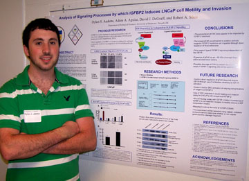 Analysis of Signaling Processes by which IGFBP2 Induces LNCaP Motility and Invasion Dylan S Audette, Adam A Aguiar, Robert A Sikes Department of Biological Sciences Although Prostate Cancer (PCa)
is easily treatable in early stages,
between 11-20% of those diagnosed have late stage and/or high grade PCa
that has metastasized to secondary tissues such as bone. Androgen
ablation therapy can temporarily improve the quality of life of a
patient. However, this eventually gives rise to an untreatable androgen
insensitive (AI) PCa, which inevitably leads to death. Over-expression
of a particular member of the IGF axis, Insulin-like Growth Factor
Binding Protein 2 (IGFBP-2), has been shown to be correlated with the
development of AI-PCa and other cancers. Using the LNCaP human prostate
cancer progression model, our lab correlated increased secretion of
IGFBP-2 with transition of (AS) PCa to (AI) PCa. Increased invasion and
motility was demonstrated using wound-healing and invasion assays in
the presence/absence of exogenous IGFBP-2. These tests were done in IGF
null conditions. IGFBP-2’s binding domains led us to theorize this was
the result of IGF1R independent signaling through integrins causing SRC
mediated phosphorylation of FAK Y397. LNCaP cells were grown in varying
concentrations of IGFBP-2 and then lysed. Analysis of phosphorylated
Focal Adhesion Kinase FAK (Y397) by western blot suggests decreasing
FAK phosphorylation as the concentration of IGFBP-2 increases compared
with positive controls. Shown here are preliminary results we will
expand in the future. We are currently engaged in studying other
signaling molecules that could also be involved in this IGFBP-2 induced
transition to an invasive phenotype including SRC and the protease
Calpain. Funding provided by the Summer Undergraduate Research Fellows
Program.
|
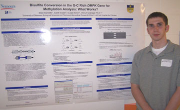 Bisulfite Conversion in the G-C Rich DMPK Gene for Methylation Analysis: What Works? Brian Barnette, Sarah Swain, Susan Kirwin, Vicky Funanage Alfred I. duPont Hospital for Children/Nemours Children's Clinic Myotonic Dystrophy Type I is an
autosomal dominant genetic disease. The disease arises from an
expansion of a trinucleotide repeat, (CTG)n located in the 3’
untranslated (3’ UTR) region of the dystrophica myotonin protein kinase
(DMPK) gene on chromosome 19. Located proximal to this region is
a CpG island; a region of DNA with a high concentration of CpG
sequences that often associate with gene promoters in humans. The
purpose of this study was to determine the methylation status of this
CpG island in individuals affected with DM1, notably congenital DM1,
and to compare this methylation pattern with that seen in
individuals not affected by DM1. The method of
bisulfite-conversion was used to ascertain the methylation status of
the CpG island. This method converts all unmethylated cytosine
residues into uracil residues. The converted DNA is then
amplified by means of PCR where the converted uracil residues are
amplified as thymine residues. This amplified DNA is then
sequenced to compare the methylation status of the CpG island among
individuals. Currently, we are working on optimizing conditions
for conversion, amplification, and sequencing of this CpG island.
These techniques will be used to explore a relation, if any, between
methylation of the CpG island and the difference in expression of
full-length DMPK transcript between adult-onset and congenital DM.
Funded by a Charles Peter White Scholarship.
|
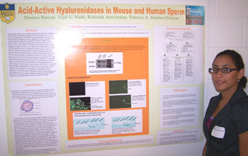 Acid-active Hyaluronidases in Mouse and Human Sperm Diniece Barran, Tejal Naik, Aravindan Rolands, and Patricia M Deleon. Department of Biological Sciences Hyaluronidases (hyases) are
membrane proteins that catalyze the
breakdown of hyaluronan, an abundant component of the extracellular
matrix of both somatic and reproductive cells. Both somatic and
reproductive hyases have been identified in mammals. In mice
reproductive hyases which play a role in sperm function include Sperm
Adhesion Molecule 1 (SPAM 1) and HYAL5, both of which have optimal
activity at neutral pH, and HYAL3 which is acidic-active. SPAM1 is
thought to be the only functional human reproductive hyase that seems
to be responsible for dispersing the cumulus cells surrounding the
oocyte, binding of the sperm to the zona on the oocyte after the
acrosome reaction and penetrating the zona due to soluble hyase
activity at pH 6. Recent data however, weaken the possibility that
either SPAM1 or HYAL5 is able to bind the zona in the sperm-egg
interaction and so it is believed that HYAL2 and HYAL3 which are
acid-active somatic hyases may play a role in the aforementioned
activities. Thus the aim of this research was to determine the function
of HYAL2 and HYAL3, acid- active hyases, in sperm function. The first
objective was to identify their presence on the sperm and the acrosomal
membranes. In both human and mouse sperm samples, Western analysis
revealed, bands for HYAL2 at a molecular weight of 54 kDa and HYAL3 at
49 kDa. Immunocytochemistry provided support for the localization of
these proteins on the sperm surface and hyaluronic acid substrate gel
electrophoresis revealed that they are functionally active. This study
is important for future hyase classification as these acid-active
hyases could be backup redundant genes for SPAM1 which is the only
reported functional human reproductive hyase.This research was
supported by the National Science Foundation, IGERT at the University
of Delaware.
|
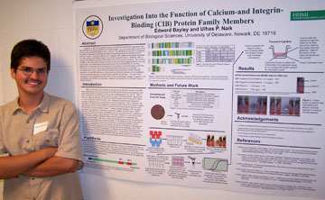 Investigation Into the Function of Calcium-and Integrin-Binding (CIB) Protein Family Members Edward B. Bayley and Ulhas P. Naik Department of Biological Science Reported in 1997, the
cytoplasmic signaling molecule now termed CIB1
was discovered and identified as a potential regulator of the platelet
integrin αIIbβ3 due to the specific nature of its binding to the
cytoplasmic domain of the αIIb subunit. Our lab has found that CIB1 is
necessary for platelet spreading on immobilized fibrinogen and does
this through its activation of focal adhesion kinase. In spite of its
specific role in platelets, CIB1 appears to be globally expressed in
human tissues and is therefore likely to have other important
functions. Moreover, CIB1 is only the first member of a family of CIB
proteins to which there are four members. The functions of CIB2, 3, and
4 are entirely unknown, and their differential expression in human
tissues suggests that they may play unique roles. Two breast carcinoma
lines were chosen (T47D and MDA-MB-231) for their difference in
invasiveness and will be analyzed for the presence of CIB family RNA by
RT-PCR. The search will continue until cell lines expressing multiple
CIB proteins are identified. To investigate their function, plasmid DNA
was prepared for the knockdown of CIB1 and CIB2 expression through RNA
interference. Cells expressing CIB family members will be transfected
with the appropriate plasmid(s) and stable clones selected with
antibiotic. The extent of the knockdown will be quantified by real-time
PCR and any morphological changes or changes in the cell motility will
be studied. Funding was provided by the Howard Hughes Medical Institute.
|
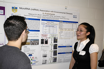 MicroRNA (miRNA) Association with Environmental Stresses in Arabidopsis Rebecca S. H. Brown, Dong-Hoon Jeong, and Pamela J. Green Department of Plant and Soil Science MicroRNAs (miRNA) are a type of
non-coding small RNA (smRNA) that
regulate gene expression at the post-transcriptional level by base
pairing with complementary sites in messenger RNA (mRNA), causing
either mRNA degradation or translational inhibition. The overall goal
of this project is to understand the relationship between miRNAs and
abiotic stresses in plants. How plants employ miRNAs to alter gene
expression when they encounter various stresses, like drought, cold,
submergence, and salinity, is of great agricultural importance. Plants
must develop sophisticated ways to cope with these stresses since they
are unable to evade them. We first utilized small RNA biogenesis
mutants such as dcl1, dcl2/3/4, or rdr2 to enrich specific small RNAs,
and then used these mutants to make smRNA libraries. We treated
Arabidopsis rdr2 seedlings with submergence, salt, and both submergence
and salt, while treating Arabidopsis rdr2 flowers with drought, salt,
and cold. Their low molecular weight RNA were isolated, and smRNA
libraries were constructed. We checked the quality of the libraries by
traditional cloning and sequencing. From these smRNA libraries, miRNA
expression will be analyzed by high-throughput sequencing by SBS
(sequencing by synthesis) and sent to a company (Illumina) or a
facility at the Delaware Biotechnology Institute to sequence, and then
for data mining by computational analysis. Potential miRNA candidates
will be identified and undergo validation. R.S.H.B was supported by
Delaware EPSCoR, through National Science Foundation Grant EPS-0447610
and the State of Delaware, and NSF grant MCB#0548569 to P.J.G. provided
research support.
|
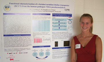 Functional characterization of a betaine/carnitine/choline transporter (BCCT) found as part of a unique cluster of systems on chromosome I of Vibrio parahaemolyticus RIMD2210633 Megan E. Carlberg, Lynn M. Naughton, and Fidelma E. Boyd Department of Biological Sciences Vibrio parahaemolyticus is a
Gram-negative halophilic marine bacterium
found in estuarine and marine coastal ecosystems worldwide. The ability
of the organism to survive and proliferate under conditions of
fluctuating salinity is well known and can be contributed in part to
the uptake of compatible solutes, organic molecules which allow the
bacterium to avoid plasmolysis due to excess water loss in a hyper
osmotic habitat. We identified a number of putative compatible solute
transporters belonging to the betaine/carnitine/choline (BCCT) family
of transporters using bioinformatic analysis on the V. parahaemolyticus
RIMD2210633 genome sequence. VP1723, a putative BCCT family transporter
was found as part of a unique cluster of systems on chromosome I of V.
parahaemolyticus. Using TMHMM, SOSUI and hydropathy profiling we
identified the transmembrane helices of this transporter and compared
our results with those of previously characterized BCCT transporters
from other organisms. We amplified VP1723 from V. parahaemolyticus and
cloned it into the broad host range vector pBBR1MCS. The vector was
then transformed into a transporter deficient E. coli strain MKH13 in
order to functionally characterize the specificity of compatible solute
uptake by this transporter. Growth of the E. coli strain under
conditions of elevated salinity will confirm a fully functional
transport system. Assays using radiolabelled compatible solutes will
determine the affinity of the transporter for betaine, carnitine or
choline. NMR analysis will allow us to determine whether uptake of
these solutes facilitates the synthesis of glycine betaine using
choline as a precursor molecule. Supported by the Charles Peter White
Undergraduate Science Education Program.
|
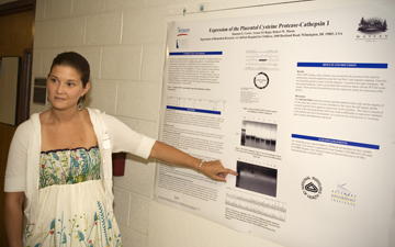 Expression of the Placental Cysteine Protease Cathepsin 1 Shannon E. Carter, Aruna Sri Bojja, and Robert W. Mason Department of Biomedical Research, A.I. duPont Hospital for Children, 1600 Rockland Road, Wilmington, DE 19803 The placenta is a highly
organized and complex organ in mammals. Its
primary function is to transport nutrients from the mother to the fetus
during gestation. The trophoblast cells that are characteristic to the
placenta are in direct contact with the maternal tissue and
consequently these cells are likely to be the key regulators of
placental function. Here the focus is on Cathepsins 1 and 2, both are
members of a family of placental cysteine proteases found on chromosome
13 in rats, and are expressed early in gestation. Both cathepsins are
localized to invasive trophoblast giant cells, and are strong
candidates as more proteolytic cathepsins involved in embryonic
nutrition or extensive remodeling during invasive implantation. The
primary objective of this 10 week study is to express cathepsin 1
respectively through such methods as RT-PCR, TOPO-cloning, RE
Digestion, and Insertion into Expression Host. Supported by Grant
Number 2 P20 RR016472-08 under the INBRE Program of the National Center
for Research Resources (NCRR), National Institutes of Health (NIH).
|
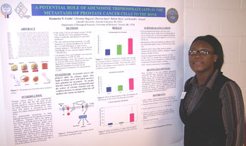 A Potential Role of Adenosine Triphosphate (ATP) in Prostate Cancer Metastasis to Bone Kiamesha N. Castle, Christine Maguire, Patricia Jones, Robert Sikes and Randall L. Duncan Department of Biological Sciences Prostate Cancer (PCa) is the
second most common type of cancer among
men and results from the uncontrolled growth of epithelial cells that
line the ducts of the prostate gland. The cause of death in these men
is rarely attributed to the original tumor, but rather the metastasis
of the cancer to its primary targets, bone and lymph. Our lab has shown
that bone cells release ATP in response to a number of stimuli. ATP
then signals surrounding cells through purinergic receptor activation
to induce bone formation. We postulate that this release may play a
role in the affinity of PCa cells to bone and that more metastatic
cells release greater amounts of ATP. To test this hypothesis, we
examined levels of ATP released from cells of the LNCaP model; LNCaP,
C4-2 and C4-2B cells. These cells were developed to become increasingly
metastatic. LNCaP cells representing the least metastatic and C4-2B
cells being the most aggressive. Our lab has demonstrated that the
release of ATP is mediated through entry of calcium into the cell. We
treated the three cell lines with ionomycin, a calcium ionophore. We
found that the C4-2B cells released the greatest amount of ATP compared
to untreated control cells and that LNCaP cells released the least.
These studies suggest that more metastatic cells release large amounts
of ATP to stimulate cell migration and invasion of PCa cells into bone.
Future studies will focus on the mechanism of ATP release from the
metastatic cells and the effector response of these cells to ATP.
(Funded by Department of Defense PCRP-W81XWH-06-1-0244 and NIH/NIAMS
R01 AR051901)
|
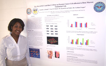 The effect of IGF-1 and RhoC GTPase on Prostate Cancer Cell adhesion to Bone Marrow Endothelial Cells Danielle A. Daniels1, Cara W. Dubyk, Kenneth van Golen, and Carlton Cooper Department of Biological Sciences, University of Delaware; 1Delaware State University RhoC is a small signaling
protein, more specifically a monomeric
GTPase, and is a member of the Ras sub family. Rho proteins are
involved in multiple cellular processes, such as cell division,
intracellular trafficking, and the organization of cytoskeletal
components. The RhoC protein has been shown to be directly involved in
cancer cell motility and invasion. Breast cancer, inflammatory breast
cancer, and pancreatic cancer are among a few of the cancers in which
RhoC is known to play an active role. Insulin-like Growth Factor
(IGF-1) plays a significant role in cell growth regulation and
development, as well as cellular DNA synthesis. This experiment uses
three different cell lines to determine their ability to adhere to Bone
marrow endothelial cells (BMEC) in the presence of IGF-1. The three
types of PC-3 cell lines used were parental PC-3, PC-3’s containing a
dominant negative RhoC (dnRhoC), and a control PC-3 vector Lac Z. These
cells were treated with IGF-1 for varying time points ranging from 5 to
30 minutes, and their adhesion to BMEC cells was measured by performing
an adhesion assay. The hypothesis of this experiment is that IGF-1
regulates PC-3 adhesion through RhoC GTPase activity. Prostate cancer
cell adhesion to Bone marrow endothelial cells is a critical step in
invasion and metastasis to bone, which is a major clinical concern.
Funding for this project has been provided by the Department of Defense.
|
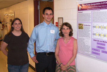 The acrB efflux pump gene is upregulated in strains of Salmonella enterica 4931 with reduced susceptibility to the quaternary ammonium compound Dodecyltrimethylammonium chloride Aleksey Dvorzhinskiy, Megan Mierswa, and Diane S. Herson Department of Biological Sciences Salmonella spp. are
gram-negative pathogens that are estimated to be
responsible for 1.4 million cases of food poisoning annually in the
United States. With the ever increasing use of anti-microbial products,
multi-drug resistant strains have appeared in natural, clinical, and
laboratory environments. This resistance is thought to be at least
partially the product of the up-regulation of membrane efflux pumps. In
this study Salmonella enterica 4931 strains were developed which were
able to grow in the presence of 500-600 ppm Dodecyltrimethylammonium
chloride (DTAC), a quaternary ammonium compound. A microarray analysis
of mRNA from a reduced susceptibility (SRS) versus a parental strain
both growing in DTAC was done. Results indicated an increase in the
amount of transcription of acrB, an efflux pump, in the SRS strain
which has been linked to multi-drug resistance. Penicillin G is another
known substrate of acrB and these new findings supported earlier
results that SRS strains with a reduced susceptibility to DTAC also
showed reduced susceptibility to Penicillin G. In addition, this study
also showed that SRS strains in the presence of the efflux pump
inhibitor Carbonyl cyanide 3-chlorophenylhydrazone (CCCP) showed a
greater sensitivity to DTAC than the parental strain. This is possibly
another physiological indication of a greater reliance of the SRS
strains on efflux pumps for defense against anti-microbial compounds.
The discovery of efflux pump inhibitors such as CCCP could lead to
advances in combating SRS strains in clinical and natural environments.
This research was generously funded by the Science and Engineering
Scholars Program.
|
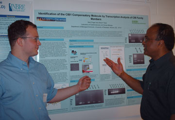 Identification of a Calcium and Integrin Binding Protein-1 (CIB1) Compensatory Molecule by RNA Expression Analysis of CIB Family Members Seth Finger and Ulhas P. Naik Department of Biological Sciences Calcium and Integrin Binding
Protein (CIB1) is primarily important in
the processes of angiogenesis, hemostatis, thrombosis and
spermatogenesis. More specifically, CIB 1-/- mice are unable to produce
sperm and exhibit defects in ischemia-induced angiogenesis. CIB1 is a
specific binding partner for Integrin aIIb β3, the platelet fibrinogen
receptor. CIB1 is a member of a family which includes three other
closely related proteins (CIB 2, 3 and 4). This study seeks to
determine if there is a member of the CIB protein family that
compensates for the absence of CIB1 and functions in way that parallels
CIB1. To test this hypothesis, RNA will be isolated from platelets,
endothelial cells, lungs and testis from wild type and CIB1-/- mice.
RNA purity will be determined by analysis of 260/280 and 260/230
absorbance ratios using a nanodrop spectrophotometer. Pure RNA samples
will be used for One Step Reverse-Transcriptase PCR utilizing specific
primers for CIB 1, 2, 3, and 4 to detect transcription of these
proteins. For each sample, a β-actin control using reverse
transcriptase and DNA polymerase will allow for the evaluation of RNA
integrity and another β-actin control without reverse transcriptase
will detect any genomic DNA contamination. In a certain tissue, if
there is a CIB family member (CIB 2, 3 or 4) that is expressed in the
CIB1-/- mouse but not in the wild type mouse, a compensatory molecule
can be identified and researched further. We have found that CIB3 is
not expressed in wild type and CIB1-/- mouse testis, whereas CIB2 and
CIB4 are both expressed equally amongst the wild type and CIB1-/-
testis. Therefore, we can conclude that CIB2 and CIB4 are not
compensating for the absence of CIB1 since no sperm are produced in
their presence. If the compensatory molecule that we identify proves to
restore function to cells that are defective in the absence of CIB1, a
new avenue of research into another CIB family member will open. This
project is supported by the Howard Hughes Medical Institute and
National Institutes of Health grant UPN.
|
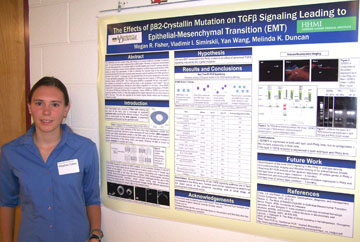 The Effects of BetaB2-Crystallin Mutation on TGFbeta Signaling Leading to Epithelial-Mesenchymal Transition (EMT) Megan Fisher, Vladimir Simirskii, Yan Wang, Melinda K. Duncan Department of Biological Sciences The mammalian eye lens consists
of fiber cells covered on the anterior surface by monolayer of
epithelial cells. The most abundant protein in adult lens fibers is
BetaB2-crystallin. Previously, our laboratory demonstrated that mice
heterozygous for a 12 nucleotide deletion in the BetaB2-crystallin gene
(Crybb2Phil) develop cataracts. However, in homozygous mutants, the
lens epithelium undergoes an epithelial mesenchymal transition (EMT)
leading to severe lens abnormalities. In order to understand the
molecular basis of this phenotype, a SuperArray real time RT-PCR gene
expression panel was used to test the hypothesis that TGFbeta signaling
is activated in the Crybb2Phil homozygous lens. We discovered
that of the 84 genes represented on the panel, 18 were significantly
upregulated in the Crybb2Phil lens indicating that some aspects of
TGFbeta signaling are induced in lenses exhibiting EMT. One of
these genes (IGFBP3) is upregulated 160 fold in the Crybb2Phil lens
epithelium as compared to wildtype, and this protein is upregulated in
the Crybb2Phil lens as assayed by immunofluorescence, although
the most notable upregulation was in the fibers. Since IGFBP3
induces EMT in other systems by binding to its receptor TGFbetaRV, we
investigated the expression of TGFbetaRV in the lens by RT-PCR and
confirmed that it is expressed. Neither IGFBP3 nor TGFbetaRV have
ever been previously reported in the lens, so these data suggest that
this receptor ligand pair may be a novel inducer of EMT in the
lens. This work was supported by the Howard Hughes Medical
Institute and the National Institutes for Health.
|
|
Localizing Compartmental Gene Expression in Urogenital Sinus (UGS) Sander B. Frank, Qian Chen, and Robert A. Sikes Department of Biological Sciences The molecular events associated
with prostate development are being
elucidated slowly. This study sought to validate the gene expression
profile from previous microarray (MA) data that showed significant
enrichment of hundreds of candidate genes in specific domains of the
urogenital sinus (UGS) namely: urogenital epithelium (UGE), urogenital
mesenchyme (UGM), urogenital dorsal (UGD), urogenital ventral (UGV)
halves. Of these candidate genes, twelve were chosen with significant
differences in both UGM/UGE and UGD/UGV, while another eleven were
chosen with significant differences in either UGE/UGM or UGD/UGV. We
collected samples of 16.5 days p.c. UGS and separated them either into
UGE and UGM, or bisected them into UGD and UGV. RNA was extracted and
used to synthesize cDNA. Oligonucleotide primers suitable for
quantitative-polymerase chain reaction were designed for each of the
candidate genes. Reaction conditions for each primer set were
determined empirically by gradient RT-PCR. Immunofluorescence (IF) was
then used to confirm protein localization for one candidate gene in
each of the eight compartments or domains of the UGS. The first round
of Q-PCR confirmed the predicted localization for 20 of 23 genes
tested, though not always at the same fold change as predicted by the
MA. The validation using IF is still ongoing, but so far has localized
clearly: Tpm in UGM, E-cad in UGE, Pax2 in UGD, Myh3 in UGM, Msc in
ventral UGM, and Fgfr2 in dorsal UGE. These techniques should result in
validation of the developmental prostate microarray results and add
critical knowledge about gene segregation, regional expression and
early morphogenetic determination in the UGS.
|
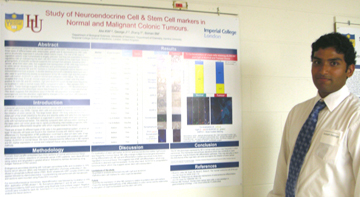 Study of Neuroendocrine and Stem Cells in Normal and Malignant Human Colonic Tissues. Joseph George1, Koree W Ahn, Tao Zhang, Bruce M Boman. 1Imperial College School of Medicine, London, United Kingdom. Christiana Care. Department of Chemistry, Hamline University. Colon carcinomas arise from the
normal epithelium of the colon. This
epithelium contains three types of differentiated cells: enterocytes,
goblet cells & neuroendocrine (NE) cells and is structurally
organized in subunits called colonic crypts. The role of NE cells in
regulation colonic crypt homeostasis is largely unknown, but they are
postulated to have a regulatory role on maintaining the stem cell (SC)
niche located at the crypt base. Since a growing body of scientific
evidence supports the concept that cancers, including colon cancer,
arise from stem cells, this suggests that dysregulation of NE cells may
contribute to colon cancer development. We qualitatively and
quantitatively compared the expression of several NE and SC markers in
normal and malignant colonic tissues using immunohistochemical
analysis. A double-labelling immunofluorescence technique was also used
to quantitatively assess co-expression of the NE marker CgA and the
putative stem cell marker ALDH1A1 in cells in normal and malignant
colon tissue. The results revealed that most, if not all, normal crypts
and colon cancers contain cells that stain positive for both NE and SC
markers. CgA and ALDH1A1 positivity was more frequently co-localized in
cells in normal crypts (90%) compared to cells in colon cancers (30%;
p<0.001). These findings suggest that NE cells frequently display a
SC-like phenotype in normal crypts but this phenotype occurs less
frequently in NE cells in colon carcinomas. This study suggests that
alterations occur in the NE cell population during colon tumorigenesis
and this alteration might lead to changes in the crypt stem cell niche
and contribute to colon cancer development.
This project was funded by Imperial College Undergraduate Research
Opportunity Programme, Christiana Care and Department of Chemistry at
Hamline University.
|
 Differential ECM Gene Expression by Parotid and Submandibular Salivary Gland Cells Kyle Green, Ben Israel, Swati Pradhan, Robert Witt, and Mary C. Farach-Carson Center for Translational Cancer Research (CTCR), Department of Biological Sciences Head and neck cancer patients
who undergo radiation therapy suffer from
salivary gland hypofunction. To help this problem, a current project is
underway that involves tissue engineering to engineer an artificial
salivary gland. As a part of this project, the purpose of this
investigation is to explore the expression pattern of extracellular
matrix genes in salivary glands. This study focuses on two of the major
salivary glands, the submandibular and the parotid. Finding the optimal
matrix will assist dissociated salivary cells to self-assemble then
differentiate into their glandular phenotype. In order to analyze ECM
gene expression, a SuperArray-based analysis of extracellular matrix
(ECM) molecules was conducted. RNA was isolated individually from
submandibular and parotid tissue and reverse transcribed to cDNA to use
in the SuperArray. The ECM gene expression of both glands was compared
and it was found that collagens, selectins, laminins, certain
integrins, MMPs, and hyluronan synthase 1 were highly expressed in the
submandibular gland relative to the parotid. Primers were designed for
these highly expressed genes and a Q-PCR-based assay was used to
validate the SuperArray results. It was found that the genes were in
fact highly expressed in the submandibular versus the parotid. Further
work will involve validating the expression of the ECM molecules at the
protein level. This research was funded by the Center for Translational
Cancer Research, the Howard Hughes Medical Institute, and by private
philanthropic contribution.
|
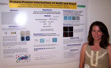 The role of protein-protein interactions in controlling Prox1 function Lauren H. Isaacs, Xiaoren Chen, Tapan P. Patel, Melinda K. Duncan Department of Biological Sciences Prox1 is a homeodomain
transcription factor that is important for
regulation of lens, liver, pancreatic, and lymphatic system
development. Although Prox1 function is understood, it is not known how
this protein is controlled. Previously, Arid2 (also called BAF200 and
zipzap) was isolated from a yeast-two-hybrid assay as a potential Prox1
interacting protein. Arid2 belongs to the ARID family of proteins that
are important for cell development, gene expression, and cell growth
regulation. It is also a vital component of SWI/SNF complexes that
function in chromatin remodeling. This work seeks to test the
hypothesis that Prox1 and Arid2 interact and to determine the
biological relevance of this interaction. A GAL4 yeast two-hybrid assay
was used to detect an interaction between proteins by the activation of
reporter genes that are transcribed if the two proteins are able to
join. Plasmids Prox1-pGBKT7 and Arid2-pACT2 were transformed into two
different yeast strains of two different mating types. These were mated
and plated on selectable media lacking nutrients that the reporter
genes are responsible for making to screen for interactions. Yeast
colonies grew on selectable media, suggesting that Prox1 and Arid2 do
interact. Fluorescent immunohistochemical analysis was also performed
on four-week C57B6 mouse lens, showing that Arid2 and Prox1 are
colocalized in the lens fiber cells. This work currently seeks to
determine if Arid2 could affect Prox1 activity within transfection
tests using the chloramphenical acetyltransferase reporter gene and our
study will continue to establish whether the interaction between Prox1
and Arid2 occurs in vivo. This research is funded by the Howard Hughes
Medical Institute.
|
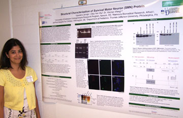 Structural Characterization of the Survival Motor Neuron Protein Upasana Joneja, Chia-Yen Wu, Wenlan Wang Departments of Biological Sciences and Chemistry and Biochemistry and the DE Nemours Biomedical Research, Alfred I. duPont Hospital for Children Spinal Muscular Atrophy (SMA)
is a neuromuscular disease characterized
by degeneration of spinal motor neurons and muscle wasting. SMA is
caused by deletion or mutation(s) in the Survival Motor Neuron 1 gene
(SMN1). The highly homologous gene, SMN2, is present in all patients
but cannot compensate for the loss of SMN1. The functions of SMN have
been implicated in the assembly of ribonucleoprotein complexes,
pre-mRNA splicing, neurite outgrowth, and cell survival. However, it
remains unclear how SMN deficiency results in selective loss of motor
neurons in SMA. The long-term goal of this project is to functionally
characterize the SMN protein. In this study, we expressed and purified
glutathione S-transferase (GST)-fused SMN protein in baculovirus-based
expression system. We cloned GST-SMN into pFastBac vector, generated
recombinant bacmid, and transfected recombinant bacmid into Sf9 cells.
We have also amplified the GST-SMN baculovirus stock and measured virus
titers by immunofluorescence staining. Small amount of GST-SMN was
purified by affinity chromatography. Western blotting analyses of the
purified GST-SMN showed accurate expression. We are currently preparing
large amount of the purified GST-SMN. We will use this recombinant
protein for structural analyses of the SMN protein by partial protease
digestion and Western blotting analyses. This research is supported by
the Howard Hughes Medical Institute's Undergraduate Science Education
Program and Dr. Wenlan Wang’s
Research Group.
|
 Allison placed third for her talk in the Sigma Xi competition. |
Identifying the potential gene targets of microRNAs involved in the bone metastasis of prostate cancer to bone Allison Kasmari, Chu Zhang, Cheng Lu, Wenzhong Wang, Blake Meyers, Pamela J. Green, and Mary C. Farach-Carson Center for Translational Cancer Research, Biology Department MicroRNAs recognize and bind to
mRNAs of target genes and alter protein
expression of those gene products.The goal of this project was to
identify and examine microRNA sequences from prostate cancer (PCa) cell
lines representing disease progression from lymph node to bone
metastases. These sequences were generated by large scale sequencing of
small RNAs found in LNCaP and its bone metastatic derivative,
C4-2B.Various bioinformatics tools and gene databases were used to
identify potential targets for both novel and known microRNAs expressed
by PCa cells during disease progression from slow growing androgen
sensitive status, represented by LNCaP, to androgen independent lethal
disease, represented by C4-2B. Prostate cancer gene expression
microarray data were used to further narrow the prospective target
genes for known microRNAs. Biobase
(http://bkl.biobase.de/cgi-bin/bkl/idb/1.0/searchengine/start.cgi)
software was used to place these gene targets in relevant cellular
pathways. Unknown potential microRNA sequences were also analyzed in an
attempt to validate their status as genuine microRNAs. Future work on
this project is designed to validate the action of microRNAs on target
genes involved in prostate cancer progression using real-time PCR.
(Supported by NCI P01 CA098912 and University of Delaware Science and
Engineering Scholars Cancer and Genetics Fellowship.)
|
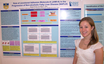 Role of junctional adhesion molecule-A (JAM-A) in the progression of non-alcoholic fatty liver disease (NAFLD) Megan Kissig and Ulhas P. Naik Department of Biological Sciences Obesity is currently the second
leading cause of preventable death in
the United States. This is due to the fact that obesity is often a
risk-factor for many diseases that eventually can cause death, such as
heart disease, diabetes, non-alcoholic fatty liver disease (NAFLD), and
others. NAFLD is characterized by the presence of an abnormal amount of
fat in the liver, which is defined as more than 10% fat by weight. My
aim was to find what effect junctional adhesion molecule-A (JAM-A) has
on weight gain and the eventual development of NAFLD. To analyze this
relationship, groups of Jam-A (+/+) and Jam-A (-/-) mice were put on
either a high fat or low fat diet for 20 weeks. During this time the
mice were weighed every two weeks to track the effects of the diet on
body weight. Every four weeks blood samples were taken with the aim of
testing for plasma levels of cholesterol and triglyceride. At the end
of the 20 weeks, the mice will be sacrificed and the livers and fat
pads will be removed to find further effects of the diet, including any
progression of NAFLD as examined through histological staining. At this
point it has been found that Jam-A (-/-) mice on the high fat diet have
gained significantly more weight than that of the other groups. This
project was supported by Grant Number 2 P20 RR016472-08 under the INBRE
Program of the National Center for Research Resources (NCRR), National
Institutes of Health (NIH).
|