Abstracts from the College of Engineering and Computer Science
Undergraduate Summer Research Symposium August 11, 2004

Ordered alphabetically by student's last name


Cloning and Expression of NK1R in Escherichia coli and Mammalian Cells
Amanda Barker3, Jason M. Winget1, and Clifford R. Robinson1, 2, 3
1Department of Chemistry and Biochemistry, 2Delaware Biotechnology Institute, 3Department of Chemical Engineering


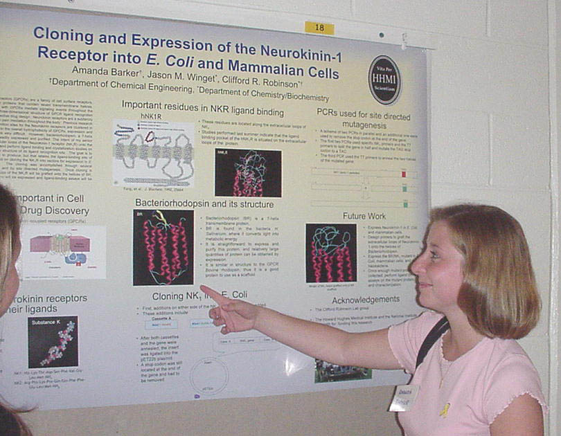
G-protein coupled receptors (GPCRs) are a family of cell surface receptors, and are integral membrane proteins that contain seven transmembrane helices.  Because ligand interactions with GPCRs mediate signaling events throughout the body, determination of the three-dimensional structure of GPCR ligand recognition sites would result in more effective drug design.  Neurokinin receptors are a subfamily of GPCRs that are involved in pain mediation throughout the body.  Previous research has shown that ligand recognition sites for the Neurokinin receptors are clustered in the extracellular loops.  Due to the overall hydrophobicity of GPCRs, expression and biophysical characterization is very difficult.  However, bacteriorhodopsin, a 7-helix transmembrane protein, is readily expressed and purified. The intent of my senior thesis is to graft the extracellular loops of the Neurokinin-1 receptor (NK1R) onto the helices of bacteriorhodopsin and perform ligand binding and crystallization studies on the mutants to determine the structure of its ligand recognition site.  The goal is to create a protein that is easy to produce, but that retains the ligand-binding site of NK1R. This summer, I focused on cloning the NK1R into vectors for expression in E. coli and mammalian cells.  The cloning was accomplished through several polymerase chain reactions, and by site directed mutagenesis.  Once cloning is complete, the extracellular loops of the NK1R will be grafted onto the helices of BR, the native and mutant proteins will be expressed and ligand-binding assays will be developed.

This project was funded in part by the Howard Hughes Medical Institute Undergraduate Science Education program.


Analysis of the Unfolded Protein Response Induced by Protein Overexpression
Elizabeth Bell, David Raden, Anne Skaja Robinson, Department of Chemical Engineering

Secretory proteins, which include proteins such as receptors, hormones, and membrane proteins, are translocated into the endoplasmic reticulum (ER), where they are folded into their active conformation with the assistance of chaperone proteins.  One important chaperone protein in the ER is BiP, which, in addition to binding unfolded proteins, binds to Ire1, preventing activation of the unfolded protein response (UPR).  The UPR is initiated when BiP dissociates from Ire1 as unfolded proteins accumulate in the ER.  The kinetics of the UPR was studied in Saccharomyces cerevisiae, budding or bakers’ yeast, induced by heterologous protein overexpression.  To easily monitor the UPR, a fluorescent stress sensor has been constructed by linking the green fluorescent protein (GFP) to the unfolded protein response element (UPRE).  The fluorescence of GFP can be measured with a fluorescent spectrophotometer, which provides a rapid readout of cell stress.  The initial kinetics of the UPR was examined along with quantitation of accumulated unfolded protein.  By extending the length of the time course beyond that of initial experiments, cyclic behavior of the UPR was observed; the mechanism of this behavior requires further study. 

This research was funded by the Howard Hughes Medical Institute Scholars Program.


3D Image Visualization of Sliced Data
Patrick Coller and Karl V. Steiner, Computer and Information Sciences and  Delaware Biotechnology Institute


This project is part of a larger program to create an interactive and immersive environment to generate and explore three-dimensional (3D) images based on biomedical imaging or microscopy-based biological data. The goal of this project is to gain a better understanding of applicable methods of interactive three-dimensional visualization. The biomedical dataset used in this project was taken from a computerized axial tomography scan (CAT scan) of an adult male torso which was obtained from the Christiana Care Hospital.  The microscopy dataset used in this project was obtained from a multiphoton confocal microscope and shows a nematode egg bursting out a root section.  In both cases the data was originally represented as a stack of individual two-dimensional grayscale slices.  It was necessary to determine if and how software and techniques used for biomedical imaging could be used to visualize microscopy data.  The raw data was first converted into specific formats that allowed it to be viewed as 3D images.  Using dedicated software packages (Amira, CaveVol and MedVol, Simian) the data could be manipulated within a intuitive, interactive 3D environment.  It was found that the hardware and software packages could be used for viewing both biomedical and microscopy data in a 3D interactive environment.

This research was supported by NIH under the BRIN grant.


Characterization of the Folding and Assembly of an Anti-Flourescein Single-Chain Antibody
Matthew DeSieno, Nathaniel Macapagal, Anne Skaja Robinson, Department of Chemical Engineering


Bacteria like Escherichia coli (E. coli) are used to produce proteins, by recombinant protein engineering techniques.  Sometimes, however, inclusion bodies form, which are inactive aggregates of the protein. The refolding of proteins from inclusion bodies using the Anti-Fluorescein Single-Chain Antibody (scFV) “4M5.3” was studied.   The process that was studied was based on denaturing, or unfolding, the inclusion body and then refolding the protein.  Previous research has found that there are four forms of the expressed protein once it has been refolded: inactive aggregate (major species), inactive monomer, and two different active monomers.  It was hypothesized that during refolding, cis/trans proline isomerization at amino acid residue 100 was the culprit for the two distinct active monomers.  The mutant V99Y (valine to tyrosine mutant at position 99) of 4M5.3 was created to favor the cis form of the two active monomers.  Once the mutant was refolded and the different forms of the expressed protein were isolated, a series of biochemical and biophysical tests were conducted in order to determine any differences between the mutant V99Y and 4M5.3.  There was little change in the distribution of active species between V99Y and 4M5.3, which suggests that cis/trans proline isomerization at residue 100 may not be the only cause for the two active forms. 

This research was funded by the Northeastern Chemical Association.


Laser Tweezer Microrheology of Gelatin
Jonathan Edwards, Chandra Sekhar Palla, and Eric M. Furst, Department of Chemical Engineering


Gelatin, a processed version of structural protein called collagen, is found in animal bones and yields a very viscous substance that transforms into a gel at high concentrations and low temperatures when prepared in water. Viscoelastic behavior of gelatin depends on the concentration, pH, solvent and thermal history of the gel. Our goal is to investigate the effects of these parameters on the structural properties of gelatin using probe microrheology. The technique involves manipulation of micron-scale particles in the gel solution with a laser tweezer. The trapped probe particle is forced in a sinusoidal motion using a laser.  Simultaneously, the response of the particle is measured with a quadrant photodiode and lock-in-amplifier.  As the particle’s motion lags behind the motion of the laser, the phase angle and amplitude of the particle are recorded and used to calculate the viscoelastic moduli. Furthermore, the dependence of these properties on temperature and concentration are also examined. 

This work is funded by the Howard Hughes Medical Institute.


The Viscoelastic Properties of MAX1 Hydrogels
Becky Gable, Cecile Veerman, and Eric M. Furst, Department of Chemical Engineering


In light of the critical shortage in donor organs used for transplants, advancements in replacement tissues developed via tissue engineering is of critical importance. Currently, artificial polymer matrices are being developed to act as an analog to the natural extracellular matrix of human tissue. One protein which could be used as a potential artificial polymer matrix is MAX1, which self-assembles, and forms a gel upon environmental changes, such as a pH change. Because these scaffolds must provide the critical preliminary molecular support that enable further development and organization of cells into a mature tissue, its proper design is crucial. The main objective of my research was to understand the mechanical properties of MAX1 via microrheological techniques. A MAX1 solution was diluted, injected with fluorescent microspheres, and its gelation process was induced by adding a pH 9 buffer. Images were then taken via video microscopy, and its gelation process was followed in time. By monitoring the Brownian motion of the particles, the microrheological responses of the MAX1 solution are currently being analyzed from the thermal motion of the fluorescent particles captured in these images, and can include statistical analysis and preliminary results.

Supported inpart by the HHMI program
.


BioExplorer – An Interactive 3D Biomedical Visualization Tool
Sungjun Kim, Department of Computer & Information Sciences
Karl V. Steiner, ECE and Delaware Biotechnology Institute


Recent software developments in simulation and 3-D visualization provide physicians and medical researchers with computer-based techniques for such applications as the education and training of medical residents, an interactive surgery planning and practice platform, or an intuitive tool to discuss certain medical conditions with patients and colleagues. This project is part of a larger research effort to develop and establish an interactive, immersive environment to explore 3D images created from biomedical or microscopic imaging methods, with the ultimate goal of developing a medical imaging simulation capacity.
The goal of this research project is to develop an interactive immersive 3D visualization tool to view and explore biomedical images. As a first step, 3D images of specific organs were extracted from a series of 2D CT scan images of a male torso, using the commercial Amira software package. For the second step, an interactive navigation program, BioExplorer, was created to enable the user to virtually fly through the 3D visualization of the torso dataset.  An interactive 3D menu was developed for the BioExplorer program that features a variety of functions such as the ability to add or remove objects in the scene, control over the objects’ resolution, transparency, and color, and interactive control over the position of several lighting sources. In a related project, work is underway to combine BioExplorer with a haptic feedback system that will include response and deformation algorithms to simulate the tactile and visual response of organs within the human body toward a medical simulation capacity.

Supported in Part by the BRIN Program.


The Role of PDI on Cellular Stress during Protein Expression
Adrienne Klotz, Ping Xu, and Anne Skaja Robinson
Department of Chemical Engineering


Secretory proteins are produced and localized using a series of compartments called the secretory pathway.  The first compartment in this pathway is the endoplasmic reticulum (ER).  The protein exits the ER when it is properly folded with the assistance of ER resident folding proteins.  When proteins do not fold they accumulate in the ER and activate an unfolded protein response (UPR) to reduce the cellular stress.  Protein Disulfide Isomerase (PDI) is an important ER resident folding assistant that has been shown to relieve the stress of a single chain antibody (scFv) expression.  PDI has three main functions:  chaperone, isomerase, and oxidase.  Mutants of PDI with combinations of these functions were studied to determine the role of PDI in relieving stress during the expression of scFv in Saccharomyces cerevisiae. The yeast cells have been engineered with a stress sensor, so that when the UPR is induced, the green fluorescent protein (GFP) will be expressed into the cytoplasm. The fluorescence of this protein can be measured by Fluorescence Spectrophotometer to indicate cell stress. To determine the role of PDI, fluorescence time courses during co-expression of scFv and mutant PDI were studied, along with analysis of intracellular scFv and PDI levels, as well as secreted scFv quantification. 

This research was funded by the Howard Hughes Medical Institute Undergraduate Science Education Program.


The Failure of Amplification of Palindromic TATA boxes
Matthew Mitsche, Josh Merritt, and Jeremy Edwards, Department of Chemical Engineering


The TATA box is a set of base pairs that precede most genes.  It is a sequence of 7 base pairs that are sequenced as TATA A/T A A/T.  When the fifth and/or seventh base pairs are altered the gene’s rate of transcription changes. .  This change is especially interesting when the TATA box is palindromic, sequenced as TATATAT.  When the sequence is a palindrome the vector will be transcribed in a seemingly random manner that has not been explained quantitatively.  The goal of my study was to insert a palindromic TATA box into vectors that have had point mutations to the gene that regulates production of glucose-6-phosphate dehydrogenase, which has been studied extensively in our lab.  After the insertion of the TATA box  the vector was transformed into yeast and the growth rate of the yeast was observed.  After the yeast growth rate was observed, a mathematical model  was constructed and compared to the growth rate of normal point mutations with the natural TATA box.  The failure came in assembling the vectors with the palindromic TATA boxes.   Sequencing  revealed that most of the G and C base pairs were eliminated so the vector that was left did not even remotely resemble the original vector, and thus,  comparison was futile.    The failure seemed to be caused by both the primers used and the inexplicable behavior of the palandromic TATA box.


Mapping Protein-Protein Contacts in β-Helical Protein Aggregates
Jessica Pippins, Mathew J. Gage, Anne Skaja Robinson, Department of Chemical Engineering


Understanding the structure of a protein aggregate provides information about aggregate formation, and can direct development of aggregation inhibitors.  However, the nature of aggregates makes structure determination by conventional means very difficult.  We are currently developing a technique to characterize the contacts between subunits within P22 tailspike aggregates using cross-linkers.  We are using the β-helix domain of the P22 tailspike protein for our studies because its production and purification are highly efficient and it has been shown to produce fibrillar aggregates resembling the aggregates involved in prion disease, Parkinson’s, and Alzheimer’s.  Using glutaraldehyde as a cross-linker, we have been able to consistently cross-link aggregated protein.  Cross-linked aggregates can then be digested using chymotrypsin, and the resulting peptide fragments are compared to peptide fragments from uncross-linked aggregates using mass spectroscopy and reversed-phase chromatography.  We hope to generate a map of the contacts with in the P22 aggregates by accumulating data from similar experiments involving different cross-linking reagents and proteases that will allow us to analyze particular locations in the aggregate structure.  This technique will hopefully be applicable to other aggregating systems.

Supported by a Science and Engineering Felowship.


Optimization of G-Protein Coupled Receptor Inclusion Body Expression in E. coli
Javier Velasquez, Steven Bane, and Anne S. Robinson; Department of Chemical Engineering


G-Protein Coupled Receptors (GPCRs) are a family of seven helical transmembrane proteins that transmit extracellular signals into the cell via association with heterotrimeric G proteins. As key players in cell signal transduction, GPCRs are involved in diseases such as cancer, HIV infection, and heart disease. They are the targets of approximately 50% of today's pharmaceuticals. Greater knowledge of the structure of these proteins would therefore be beneficial in the advancement of drug treatments. However, current overexpression systems produce GPCRs in low abundance and milligram amounts are necessary before detailed structure studies are possible. It is the goal of this project to express GPCRs at sufficient quantities in inclusion bodies within E. coli that they may be purified and refolded for structural studies. To this end, four GPCRs (mouse and human neurokinase and the human adenosine receptors A2a and A3 ) were compared in a number of E. coli cell lines. Growing temperature, induction agent levels and growth times were also studied. Expression efficiency was determined by gel electrophoresis with Coomassie Brilliant Blue stain, and confirmed by western blot assay. 

This research was funded in part by the HHMI Undergraduate Science Education Program.


Optimization and Characterization of G-Protein Coupled Receptor Expression in Yeast
Alison Wedekind, Ronald T. Niebauer, Anne Skaja Robinson, Department of Chemical Engineering

Adenosine receptors are membrane proteins that play an important role in cell signaling and response through their interactions with adenosine.  The adenosine receptors are a sub-family of the G protein-coupled receptor (GPCR) super-family.  GPCRs, which mediate cellular responses to diverse stimuli, have been implicated in many human diseases such as heart disease and asthma and are important drug targets.  Knowledge about GPCR structure and function is limited by the difficulties associated with producing large amounts of functional protein.  Previous experiments have shown that when one of the adenosine receptors, A2a, is expressed in yeast, a characteristic decrease in protein production occurs over time.  Thus, one of the research goals has been to gain insight into the causes of this decrease and create an optimal system for A2a protein expression.  For these studies, the A2a protein has been genetically fused to a green fluorescent protein (GFP), so that expression and trafficking may be monitored by fluorescence.  Recent research has included the use of a cDNA library and a flow cytometry screening technique to identify proteins that may help improve A2a production.  Other experiments have focused on improving the strength of the galactose promoter which plays a critical role in A2a expression.  Future work with A2a expression includes optimizing variables such as media pH and expression temperature and monitoring their effect on total and functional protein levels.

Funded in part by the HHMI program.


Links: Summer 2004 Undergraduate Research Symposium, Symposium Abstracts from other Colleges and Departments,
Undergraduate Research Summer Enrichment ProgramUnversity of Delaware Undergraduate Research Program, Howard Hughes Undergraduate Program.
Created  2 August 2004. Last up dated 13 August 2004 by Hal White
Copyright 2004, University of Delaware