Abstracts from the Department of Chemistry and Biochemistry
Undergraduate Summer Research Symposium August 8, 2002

Ordered alphabetically by student's last name


Synthesis and Characterization of Phosphines 
as Novel Substrates of Sulfhydral Oxidase

Steve Brohawn, James Psathas, and Colin Thorpe
Department of Chemistry and Biochemistry
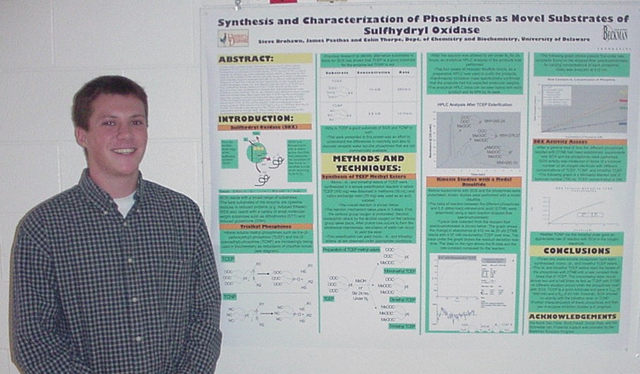
Commercially available trialkyl phosphines such as tris-(2-carboxyethyl)-phosphine (TCEP) and tris-(2-cyanoethyl)-phosphine (TCNP) are used in biochemistry as reductants of disulfide bonds. TCEP, but not TCNP, is a good substrate for chicken egg white sulfhydral oxidase (SOX), a flavoenzyme that catalyzes the oxidation of free thiols to disulfide bonds in a variety of substrates while reducing oxygen to hydrogen peroxide. In an effort to understand these differences in reactivity with the enzyme and possibly to find versatile water soluble phosphines, mono-, di-, and trimethyl ester derivatives of TCEP have been synthesized. Kinetic studies of TCEP, TCNP, and the methyl ester derivatives have been performed on a stopped flow spectrophotometer using DTNB as a model disulfide. The di- and trimethyl ester derivatives have a second order rate constant three times higher than that of TCEP and the monomethyl esters rate constant is close to two and a half times higher than that of TCEP. TCNP was found to be much less reactive than TCEP or its derivatives. SOX activity with TCEP, TCNP, and the trimethyl ester was compared using an oxygen electrode. SOX activity with TCEP gave a Vmax of 1650/min and a Km of 43 mM. The trimethyl ester derivative, like TCNP, was found to have undetectable activity towards SOX. These studies will form the basis for the investigation of the inhibition of SOX with metal ions.
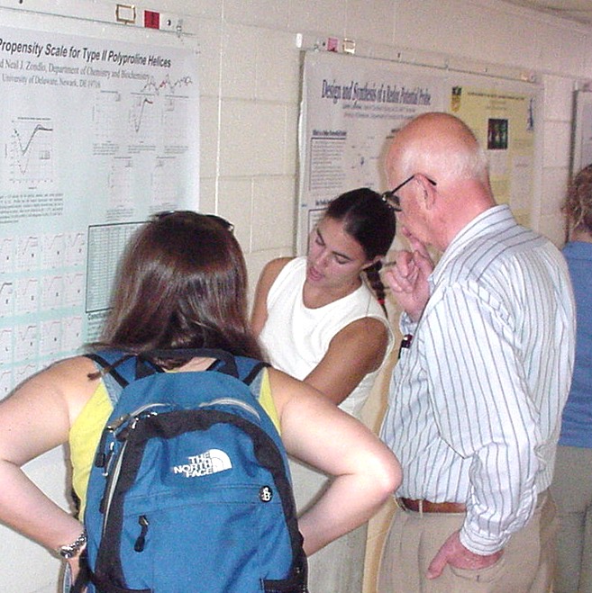

 
 
Thermodynamic Propensity Scale for Type II Polyproline Helices 

Alaina M. Brown and Neal J. Zondlo
Department of Chemistry and Biochemistry

Although type II polyproline (PPII) helices and proline-rich peptides are common structures in globular proteins, little is known about their stability, dynamics, and fundamental energetics. The ability to predict regions of high propensity for polyproline II helices is crucial to the analysis of intermolecular and intramolecular interactions mediated by PPII helices as well as predictions of possible signaling motifs.  Sequences of three or more consecutive prolines induce PPII helix formation.  A model peptide (Ac-GPPXPPGY-NH2) was designed with two proline residues on each side of a randomized position, X.  Peptides with each of the twenty amino acids in the randomized position were tested using circular dichroism (CD) spectroscopy to determine the relative stabilization effect on propagation of a PPII helix.  The CD signal of each peptide was compared to the CD signal of the reference peptide (X=P) at 229 nm to determine the relative PPII helix stability.  Beta branched amino acids (deltadeltaG~1.6 kcal/mol), cysteine, serine, histidine, and asparagine (deltadeltaG~1.0 kcal/mol), were all found to have a significant destabilizing effect on the PPII helix, while all other amino acids were comparable to the reference peptide.  Helicity was temperature-dependant and pH-dependant for charged residues, but independent of salt and peptide concentration.

\
 
 
Substrate Assisted Protein Folding 
in Pseudouridine Synthase Mutants

Christopher Eller and Eugene Mueller, 
Department of Chemistry and Biochemistry

Pseudouridine synthases catalyze the conversion of uridine to pseudouridine (PseudoU) in RNA.  Four families of Pseudouridine synthases have been identified, represented by TruA, TruB, RluA, and RsuA in Escherichia coli.  While homologous, the four families have no statistically significant sequence similarity.  A single aspartic acid residue is the only absolutely conserved residue, and it is essential for catalysis [Ramamurthy V., Swann, S.L., Paulson, J.L., Spedaliere, C.J., and Mueller, E.G. (1999) J. Biol. Chem. 274, 22225-22230].  Three of the families share a short sequence motif that contains adjacent lysine and proline residues.  Mutagenesis of the conserved lysine and proline residues in TruB and RluA resulted in proteins with circular dichroism (CD) spectra of significantly reduced intensities, suggesting a loss of secondary structure associated with protein unfolding.    The lysine and proline mutants are less stable, as indicated by storage behavior and a decreased melting point.  Despite the apparent loss of structure, the catalytic activity of these altered enzymes was only mildly impaired [Spedaliere, C.J., Hamilton, C.S., and Mueller, E.G. (2000) Biochemistry 39, 9459-9465]. In light of these observations, CD spectra and melting curves were recorded for both wild-type TruB and the P20G mutant in the presence of a 17-mer RNA substrate, the T-arm stem loop of tRNAPhe, to determine if RNA binding to the unfolded protein stabilizes the native conformation, which will be detected as an increase in the CD spectra intensity and the melting temperature.


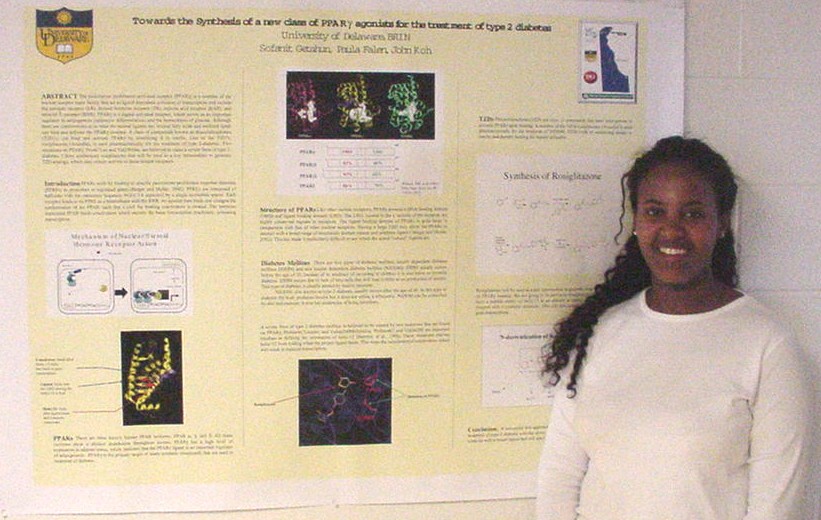
Towards the Synthesis of a New Class of PPARgamma Agonists for the Treatment of Type 2 Diabetes

Sofanit Getahun1,2, Paula Falen2, and John Koh2
1Delaware Tech and 2Department of Chemistry and Biochemistry, University of Delaware

The peroxisome proliferator-activated receptor (PPARgamma) is a member of the nuclear receptor super family that act as ligand dependent activators of transcription and include the estrogen receptor (ER), thyroid hormone receptor (TR), retinoic acid receptor (RAR), and retinoid X receptor (RXR). PPARgamma is a ligand-activated receptor, which serves as an important regulator in adipogenesis (adipocyte differentiation) and the homeostasis of glucose. Although there are controversies as to what the natural ligands are, several fatty acids and oxidized lipids can bind and activate the PPARgamma receptor. A class of compounds known as thiazolidinediones (TZD’s) can bind and activate PPARgamma by sensitizing it to insulin. One of the TZD’s, rosiglitazone (Avandia), is used pharmaceutically for the treatment of type 2-diabetes. Two mutations on PPARgamma, Pro467Leu and Val290Met, are believed to cause a severe form of type 2-diabetes. I have synthesized rosiglitazone that will be used as a key intermediate to generate TZD analogs, which may restore activity to these mutant receptors.


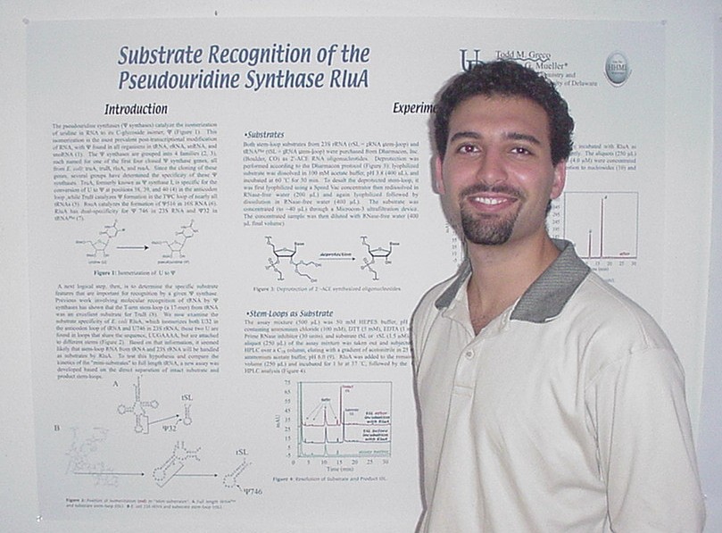
Substrate Recognition of the Pseudouridine Synthase RluA

Todd M. Greco and Eugene Mueller 
Department of Chemistry and Biochemistry

The pseudouridine synthases  catalyze the isomerization of uridine in RNA to its C-glycoside isomer.  This isomerization is the most prevalent post-transcriptional modification of RNA, with pseudouridine being found in all organisms, occurring in tRNA, rRNA, snRNA, and snoRNA [Grosjean, H. and Benne, R. (1998) Modification and Editing of RNA, ASM Press, Washington, DC].  Previous work involving molecular recognition of tRNA by pseudouridine synthases has shown that the T-arm stem-loop (a 17-mer) from E. coli tRNA­ was an excellent substrate for TruB [Santi, D.V., Gu, X., Yu, M., and Ivanetich, K. (1998) Biochemistry 37, 339-343].  We now examine the substrate specificity of E. coli RluA that isomerizes both U32 in the anticodon loop of tRNA and U746 in 23S rRNA, which are found in identical loop sequences (UUGAAAA) but have different stem sequences.  When RluA was incubated with the appropriate stem loops from tRNAPhe and 23S rRNA, total digestion and HPLC analysis showed that pseudouridine was formed.  In the case of the anticodon stem-loop, the intact product stem-loop (with pseudouridine) was resolved from substrate (with U).  The analysis showed quantitative conversion to product.  A new assay was developed to utilize this separation of intact substrate and product stem-loop to compare the kinetics of the "mini-substrates" to full length tRNA.


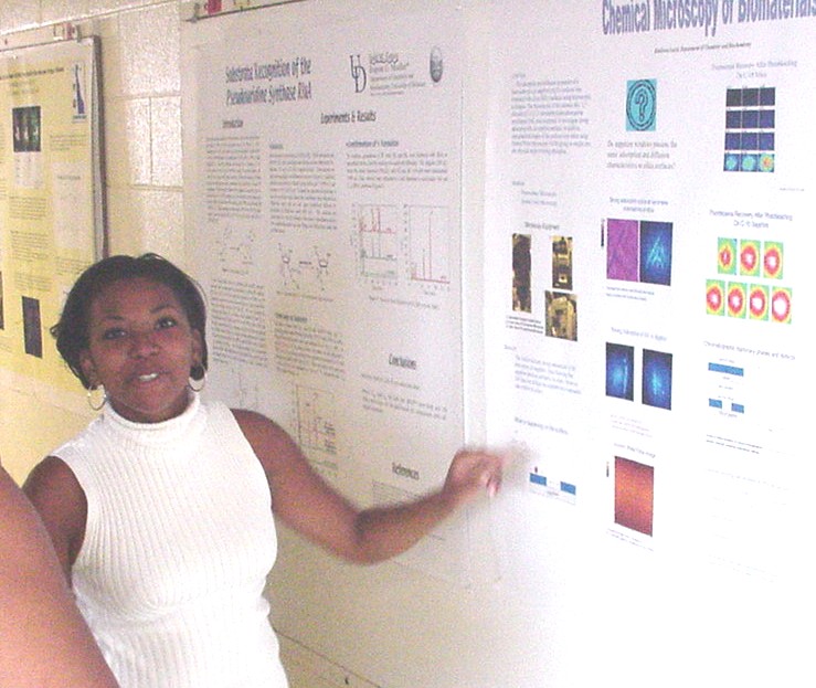
Chemical Microscopy of Biomaterials

Kholiswa Laird, Melody Ludes, Derrick Swinton, Mary J Wirth
Department of Chemistry and Biochemistry

Adsorption and diffusion of a fluorescent dye on sapphire (Al2O3) surfaces was compared with silica (SiO2) surfaces using microscopic techniques. Fluorescence of the cationic dye, 1,1’-dioctadecyl-3,3,3’,3’ -tetramethylindocarbocyanine perchlorate (DiI), was used to investigate strong adsorption sites on sapphire surfaces using an upright microscope and an ICCD camera for imaging. In addition, topographical images of the surfaces were taken using Atomic Force Microscopy (AFM) giving us insight into the physical origin of strong adsorption. The results indicate strong adsorption of DiI does occur on sapphire, thus showing that sapphire behaves similarly to silica. However, DiI does not diffuse on sapphire at a reasonable rate relative to silica.


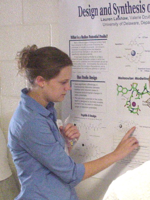
Design and Synthesis of a Redox Potential Probe

Lauren Latshaw, Valerie Dzubeck, Kyung Lee, and Joel P. Schneider
Department of Chemistry and Biochemistry

The purpose of this research is to synthesize and elucidate the properties of a tunable reduction potential probe.  The probe consists of a chromophore (carbostyril), a hexapeptide (sequence –CVDPGTC-), and a metal chelate (LnDTPA).  This probe links sensitized emission to a disulfide bond formation event.  The cysteines will form a disulfide bond in an oxidizing environment causing the formation of a ?-turn in the peptide chain portion of the probe. In this oxidized conformation, the metal chelate and the chromophore are brought into close spatial proximity and sensitized emission occurs.  Under reducing conditions, the disulfide bond is reduced and the probe adopts a conformation in which the metal chelate and chromophore are spatially far apart, thus inhibiting sensitized emission.  The probe is synthesized convergently: a DTPA pentapeptide and a carbostyril/cysteine compound are covalently joined using a segment condensation reaction.  The probe will then be side chain deprotected, oxidized, and terbium chelated.  Once the probe is prepared, its reduction potential will be measured using fluorescence.


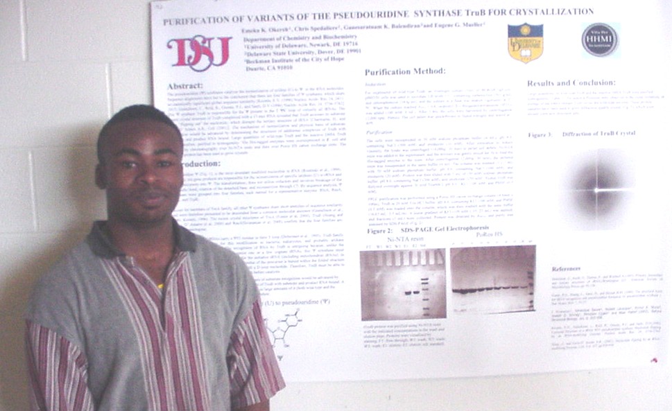
Purification of Variants of the Pseudouridine Synthase TruB 
for Crystallization

Emeka K. Okereh1,2, Chris Spedaliere2, Ganesaratnam K. Balendiran3, and Eugene G. Mueller2
1Delaware State University, 2Department of Chemistry and Biochemistry University of Delaware
3Buckman Institute of the City of Hope Duarte, CA 91010

The pseudouridine synthases catalyze the isomerization of uridine (U) to pseudouridine  in the RNA molecules. Sequence alignments have led to the conclusion that there are four families of pseudouridine synthases, which share no statistically significant global sequence similarity [Koonin, E.V. (1996) Nucleic Acids. Res. 24, 2411-2415; Gustafsson, C., Reid, R., Greene, P.J., and Santi, D.V (1996) Nucleic Acids Res. 24, 3756-3762].. The pseudouridine synthase TruB is responsible for the pseudouridine  present in the T-pseudouridine-C loop of virtually all tRNAs. The mean crystal structure of TruB complexed with a 17-mer RNA revealed that TruB accesses its substrate U by "flipping out" the nucleotide, which disrupts the tertiary structure of tRNA [Charmaine, H., and Ferre-D' Amare A.R., Cell (2001)]. The mechanism of isomerization and physical basis of substrate recognition would be advanced by determining the structures of additional complexes of TruB with substrate and product RNA bound. Large quantities of wild-type TruB and the inactive D48A TruB were, therefore, purified to homogeneity. The His-tagged enzymes were overexpressed in E. coli and purified by chromatography over Ni-NTA resin and then over Poros HS cation exchange resin. The purified protein has been used to grow crystals.


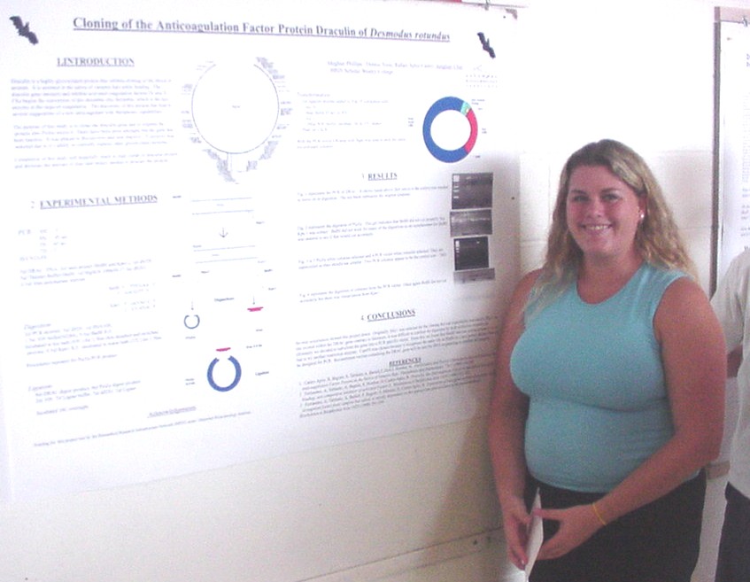
Cloning of the Anticoagulation Factor Protein, Draculin, of the Vampire Bat, Desmodus rotundus

Meghan Phillips1,2, Rafael Apitz-Castro2, Dennis Yoon2, and Jungheui Chen2
1BRIN Scholar from Wesley College and 2Department of Chemistry and Biochemistry, University of Delaware

Draculin is a highly glycosylated protein which acts as an inhibitor against blood clotting.  It is found in the saliva of vampire bats (Desmodus rotundus) and is secreted during feeding.  Previous studies have shown that this protein interacts and inhibits coagulation factors IX and X.  The discovery of this protein has lead to several suggestions of a new anticoagulant with therapeutic capabilities.  The purpose of this study is to clone the draculin gene and express the protein in a special type of yeast.  Post-translational modifications, in particular glycosylation, are difficult to confirm by expression in typical E. coli or Baculovirus expression systems.  Therefore, the yeast used in this study is Pichia pastoris.  P. pastoris has been shown to express correctly in other glycosylated proteins.  The AOX promoter is used to induce expression of the gene, which normally codes for a protein involved in methanol metabolism.   One feature of this yeast is it can survive with methanol as its sole carbon source.  Once the draculin gene is expressed and is found to be active in this yeast, further study of this protein will be conducted to determine its potential as a therapeutic agent.


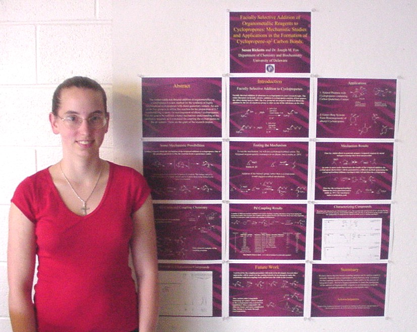
Facially Selective Addition of Organometallic Reagents to Cyclopropenes: Mechanistic Studies and Applications in the Formation of Cyclopropene-sp2 Carbon Bonds.

Susan Ricketts and Joseph M. Fox, 
Department of Chemistry and Biochemistry

The copper-catalyzed directed addition of organometallics to cyclopropenes is a new method for the synthesis of highly functionalied cyclopropenes with chiral quaternary centers.  An aim of the Fox group is to utilize this reaction for the preparation of 5-7 membered ring systems via rearrangement of alkenyl cyclopropanes.  For this goal to be realized, a better mechanistic understanding of the addition is required, as is a protocol for coupling the cyclopropane to sp2 centers.  These are the goals of the research project.


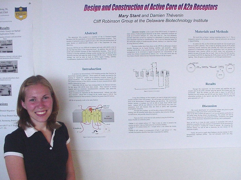
Design and Construction of Active Core of A2a Receptors

Mary Stant, Damien Thévenin, and Cliff Robinson 
Department of Chemistry and Biochemistry and the Delaware Biotechnology Institute

The adenosine A2a receptor is a member of the G-protein coupled receptor  (GPCR) superfamily of seven-helix transmembrane receptors. This receptor is  believed to play a significant part in the onset and advancement of many factors of cardiovascular disease, and is thus important for drug discovery. Many drug development and pharmaceutical research programs are interested in the mechanism of A2a receptor functions in order to develop medical and other therapeutic agents that target this class of receptors.  However, GPCRs are very difficult to express and work with which is due to the fact that they have a low expression level.  In addition, they are hard to purify and crystallize because of their hydrophobic domains.  Thus, we are working to develop methods to design smaller analogs that can keep the key functions and that will make the study of these GPCRs easier.  Through these analogues, we will be able to look at GPCR ligand binding, stability, crystallization, biophysical studies, and uses for drug discovery.


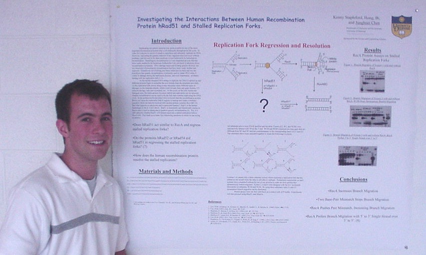
Investigating the Interactions of Human Recombination Protein hRad51 
with Stalled Replication Forks.

Kenny Stapleford, Hong Bi, and Junghuei Chen, 
Department of Chemistry and Biochemistry

Homologous recombination in both prokaryotes and eukaryotes is used for a variety of reason in the cell with one of the most important uses being DNA damage repair.  Scientist are now turning to another form of damage that can occur, and this involves possibly one of the most important biochemical processes in the cell, replication.  It is found that in Escherichia Coli, replication forks can become inactivated by DNA damage on the template strand, causing replication to stall.  Recombination proteins; RecA, RecG, and RuvAB, can then regress the fork and fix the problem.  The human homologue to RecA is the human recombination protein hRad51, and the question is now whether hRad51 can be used to fix these stalled replication forks or are other proteins such as hRad52 or hRad54 needed to allow the cell to continue replication.  We are testing these questions by using a series of six synthetic replication forks designed to represent stalled replication forks in the cell.  RecA was used in preliminary experiments as a control and hRad51 will then be used in the future to determine if it does indeed promote the resolution of the stalled replication forks.


Selective Enone Reductions Using TADDOL Ligands with High Enantomeric Control

Benjamin A. Thuma and Douglass F. Taber. 
Department of Chemistry and Biochemistry

The development of highly enantioselective reducing processes for conjugated carbonyls into their corresponding alcohols is of great importance in biological related synthesis.  The use of chiral ligands have been shown to allyate aldehydes using chiral bidentate titanium(IV) complexes with high control of absolute configuration of the resulting alcohol 3.  The use of a, a, a’, a’-Tetraaryl-2, 2-dimethyl-1, 3-dioxolan-4, 5-dimethanol (TADDOL) have been shown to be a highly effective moiety for production of the necessary reducing complex using LiAlH4 for the enantiomericly controlled reductions of ketones 4.  The reductions of ketones using the "TADDOL" complex showed an enantiomer ratio of up to 96:4 of the (S) configuration 4. Our research has shown that the “TADDOL” reducing complex was able to reduce 1,1-dimethylethyl 7-oxo-phenylheptanoate to its corresponding conjugated alcohol with an enantiomeric excess of 77.5% for the (S) configuration at -78°C. We hope to conduct further research on the  “TADDOL” reducing complex to increase the enantiomeric excess of the conjugated alcohol of 1,1-dimethylethyl 7-oxo-phenylheptanoate and of the selective reductions of long chain enones.


Links: Summer 2002 Undergraduate Research Symposium, Symposium Abstracts from other Colleges and Departments,
Undergraduate Research Summer Enrichment ProgramUnversity of Delaware Undergraduate Research Program, Howard Hughes Undergraduate Program.
Created 3 August 2002. Last up dated 12 August 2002 by Hal White
Copyright 2002, University of Delaware