X-Ray Tomography of Vascular Corrosion Casts
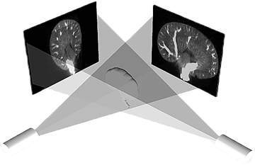 |
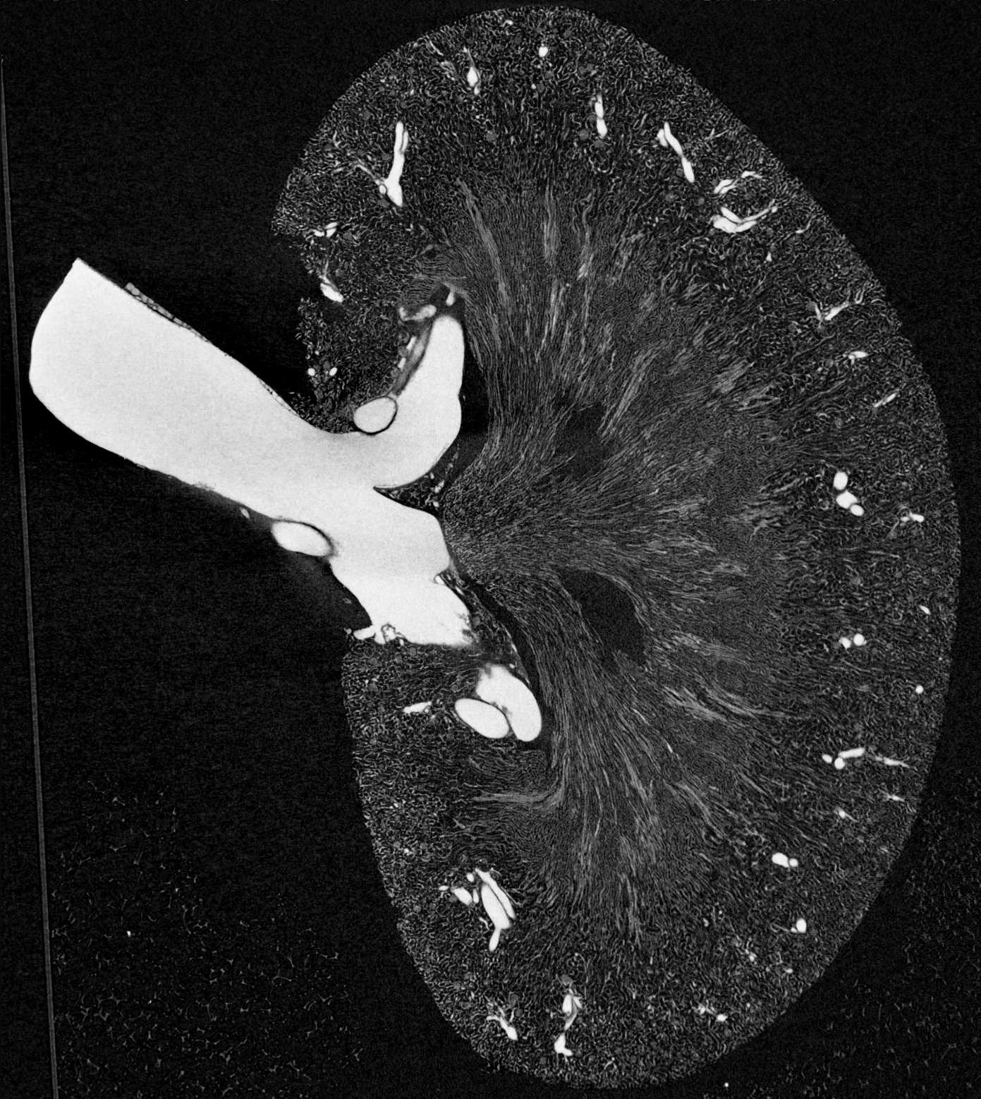
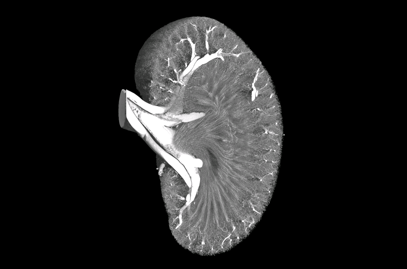
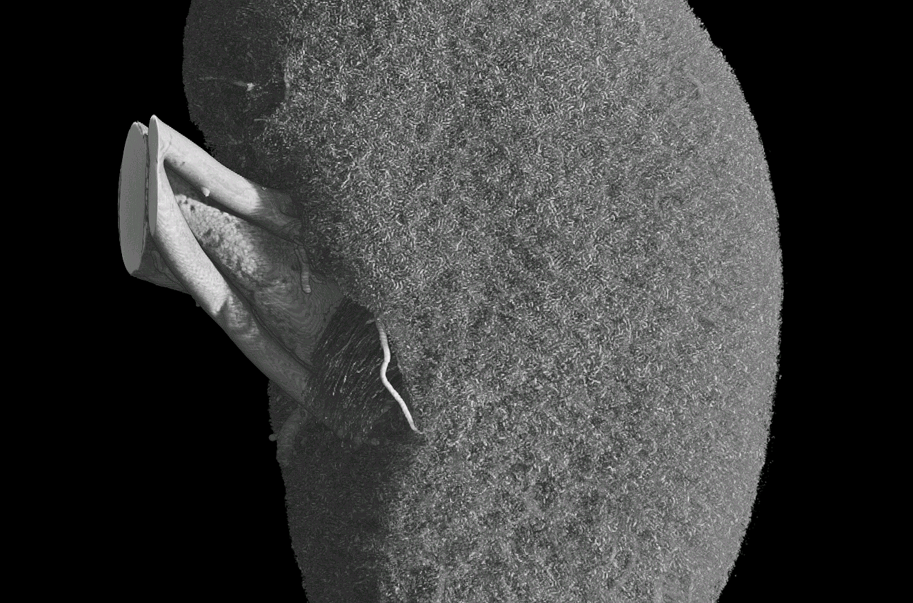
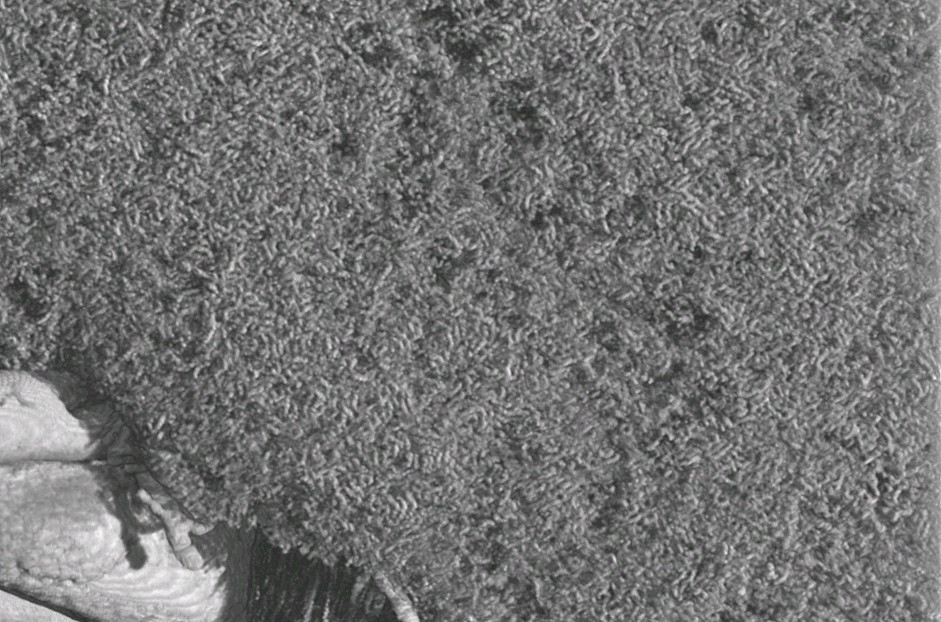
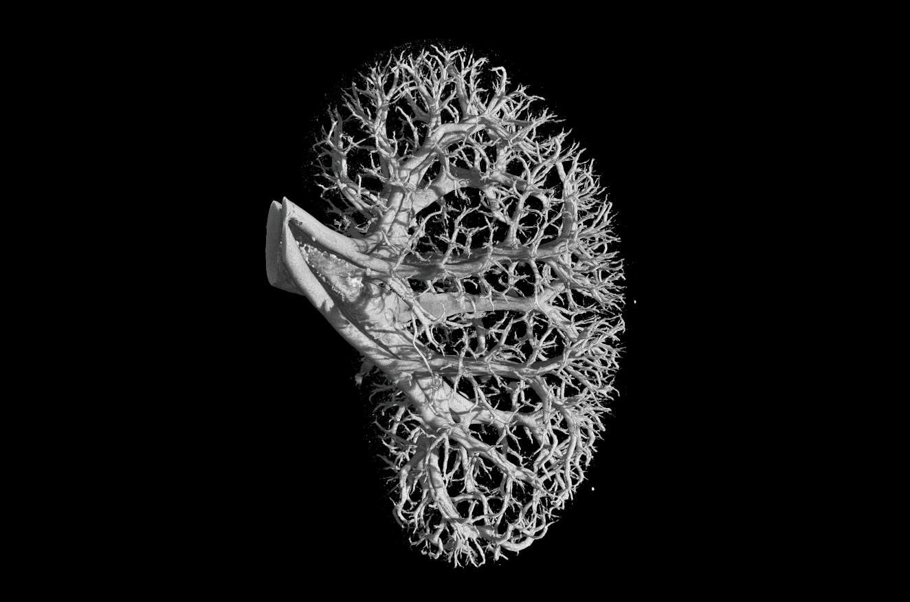
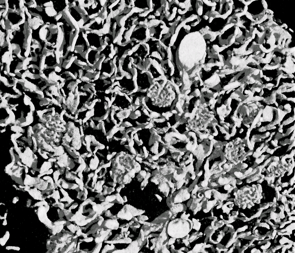
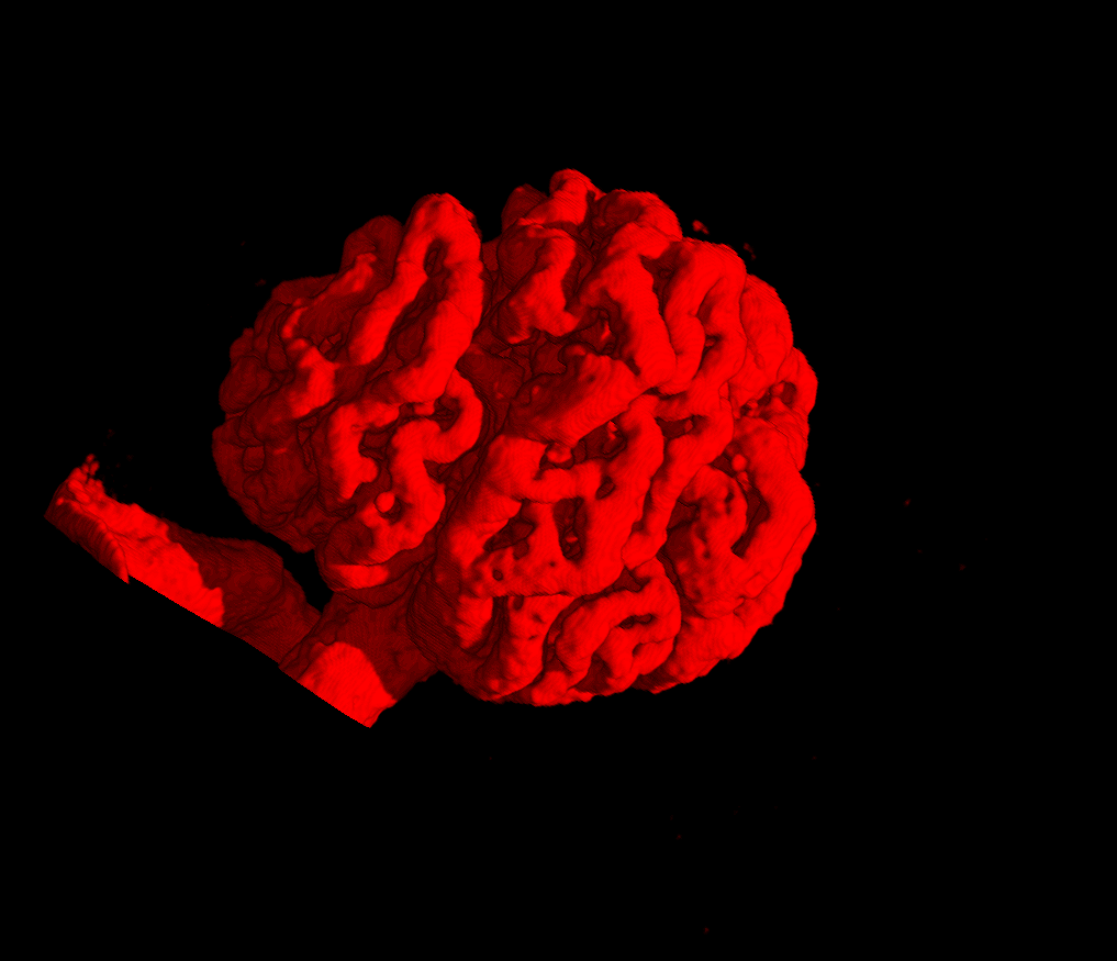
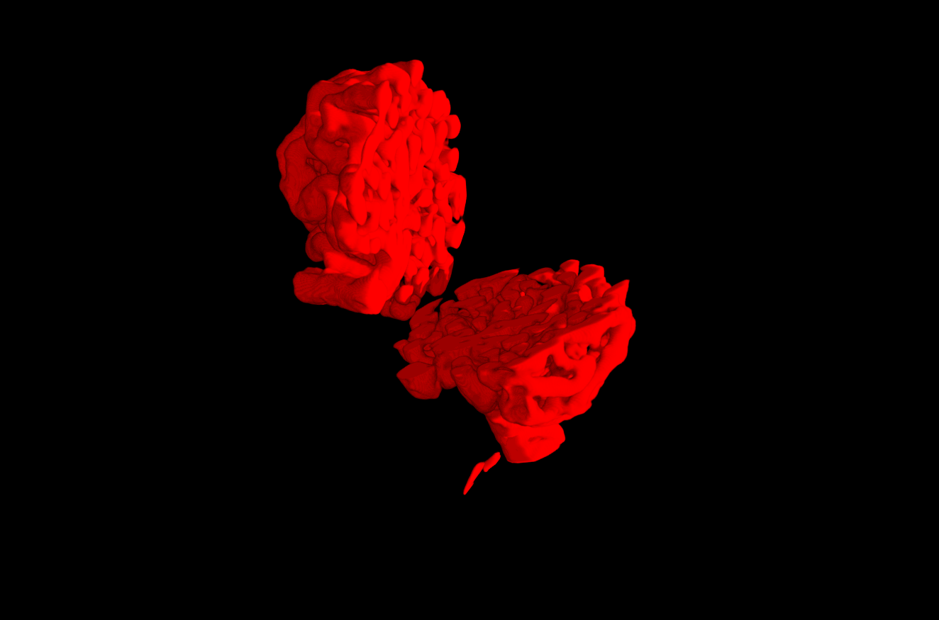
The field of view of a confocal microscope is very limited and only small portions of corrosion casts (i.e. kidney glomeruli) of larger vascular systems can be acquired as three-dimensional data sets. Larger corrosion casted specimens (whole organs and organisms) can be viewed with a Micro-or Nano-ct which use x-rays to image dense materials. The casting plastic used for corrosion casting is x-ray dense and can be made even more so by adding a lead compound to the unpolymerized resin. X-rays projected through a specimen are impeded by x-ray dense regions. X-ray projections through the entire specimen are collected tomographically by scanning the specimen in a circular pattern throughout its depth.A three dimenional data set is thus acquired with casts represented as x-ray dense regions (the images are usually reversed so that these regions appear light against a black background). Three dimensional processing program such as Amira can then be used to render the surface of the casts and provide analytical and viewing options which permit the examination of the models from any angle and also from within. The nanoct data and models below were prepared by Dr. Denis Van Loo, UGCT, Center for X-ray Tomography of Ghent University Proeftuinsrtaat 86 , 9000 Ghent, Belgium.
 |
 |
 |
 |
 |
 |
 |
 |
 |
 |