 |
Scanning Electron Microscopy of Corrosion Casts
 |
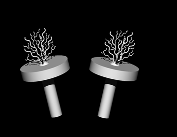
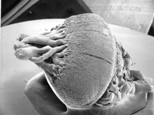
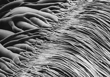
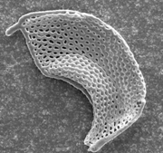
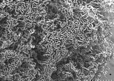
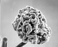
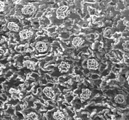
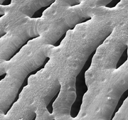
Scanning electron microscopy (SEM) of corrosion casts provides excellent three-dimensional views of the internal surface and geometry of blood vascular systems. SEM images have remarkable depth and tilting the specimen can reveal additional vessel relationships, However, upon tilting, relationships and relative dimensions change in a non-linear fashion due to parallax. This makes it difficult to obtain reliable quantitative data. In addition, the interior organization of compact vessel systems, such as the glomerulus of the kidney, remain hidden using this surface-imaging approach. The images below illustrate the sharpness and clarity of SEM images of selected corrosion casts. The Corrosion Casts and Sems were prepared by Dr. Fred Hossler, Dept. of Anatomy and Cell Biology, J. H. Quillen College of Medicine, East Tennessee State University College of Medicine, John City, Tennesse. Each image is linked to a larger version.
 |
 |
 |
|
 |
 |
||
 |
|||
 |
|||
video of tilting of glomeulus cast in the SEM |
choriocapillaries of frog eye | retinal capillaries of frog eye |