Vascular Corrosion Casting
 |
Vascular Corrosion Casting
|
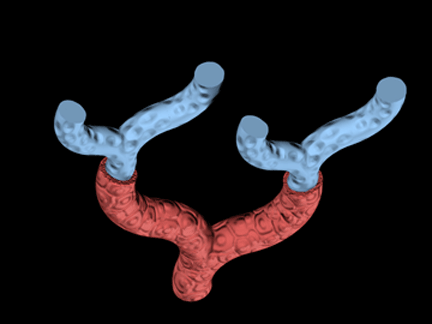
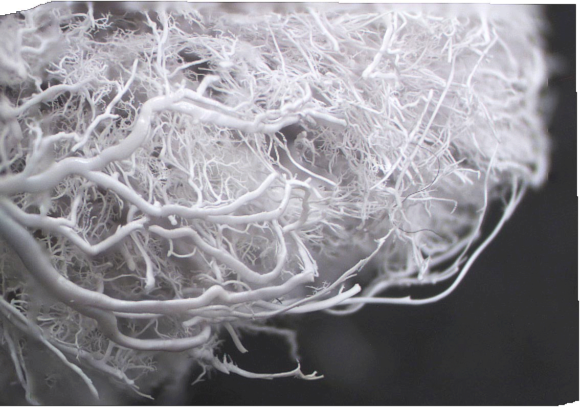
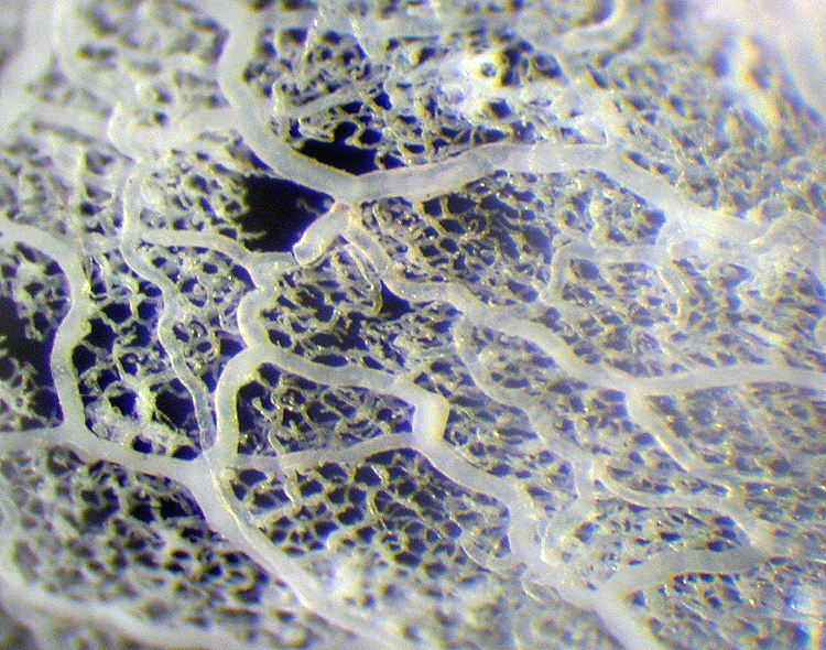
The entire vascular system of a whole animal or organ down to the smallest capillaries can be injected with a plastic polymer which fills the blood space and rapidly polymerizes. When the tissue is subsequently corroded away with an alkali solution, a hardened cast remains which accurately represents the essential geometry of the original vascular system. These casts can be examined with dissecting microscope (with limited magnification and depth of field) or a scanning electron microscope which provides striking resolution and depth of focus. Examples are shown below. These corrosion casts were prepared by Dr. Fred Hossler, Dept. of Anatomy and Cell Biology, J.H. Quillen College of Medicine, East Tennessee State University, Johnson City, Tennessee. Click on an image to access a larger version.
 |
 |
|
| Rabbit ovary photographed with light microscopy. | Mouse paw corrosion cast photographed with light microscopy,. | Corrosion cast of microvessels in the prostate gland-light microscopy. |
 |
 |
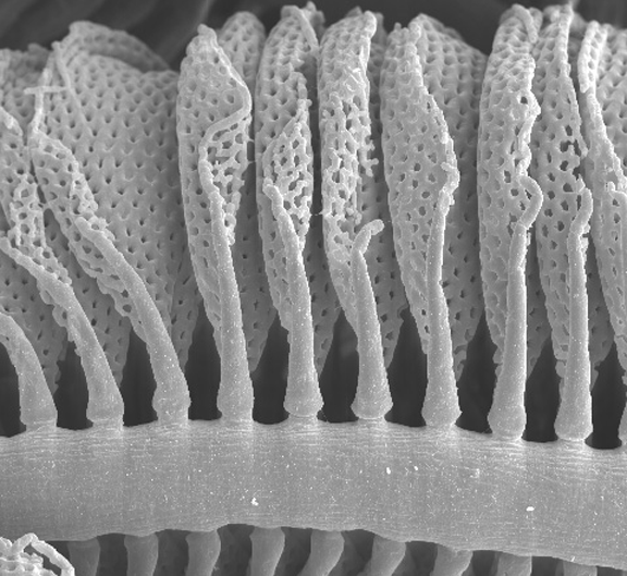 |
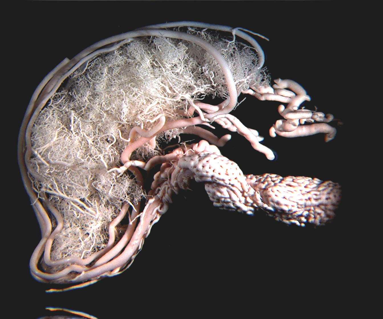
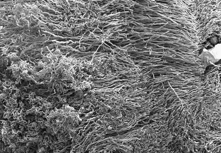
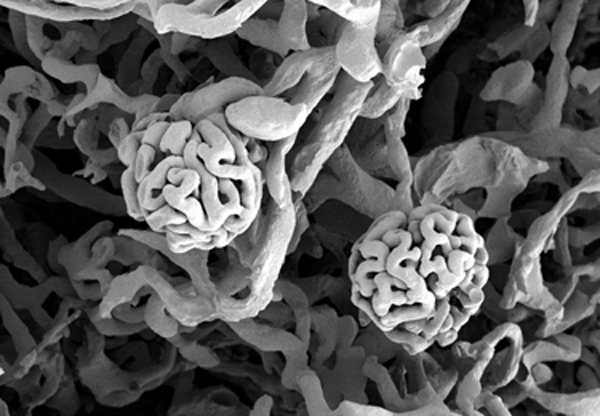
| Corrosion cast of kidney microvessels-scanning electron microscopy. | Corrosion cast of two glomerular capillary bundles in the kidney-scanning electron micrograph. | Corrosion cast of capillaries on gill filaments of a fish-scanning electron micrograph. |