Confocal Microscopy of Corrosion Casts
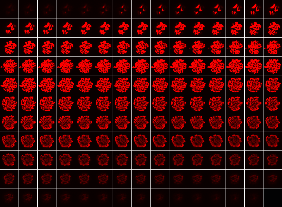
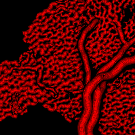
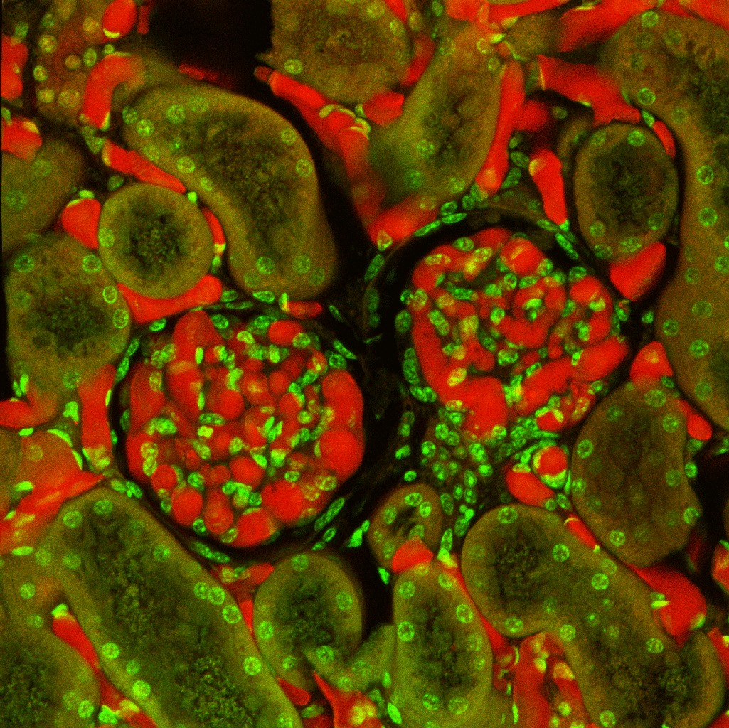
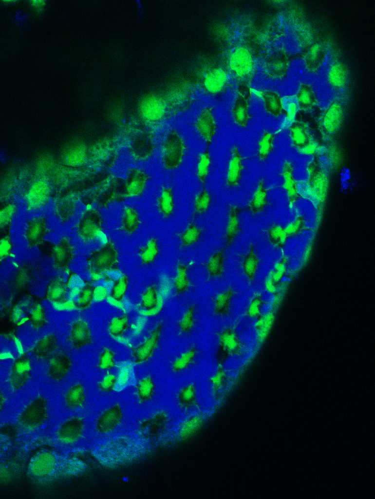
The casting polymer from which a corrosion cast is made has fluorescent properties, that is, when excited by a light beam of a specific wavelength, it emits light at a longer wavelength. A corrosion cast, therefore is an ideal specimen for examination by confocal microsopy. In laser confocal microscopy (LSM) , a laser beam excites fluorescent substances by scanning across a specimen in a square pattern called a raster. The resulting fluorescent signal is collected from only a single focal plane. This results in sharp, high contrast fluorescent images due to elimination of fluorescence above and below the focal plane. Successive optical sections can be acquired by moving the focus in increments through the specimen, a process known as optical sectioning. Numerous optical sections through through the specimen are known as a z-stack and results in a three-dimensional data set which whichiis stored digitally as voxels, a cubic picture units analagous to pixels. The casting polymer emits a strong fluorescnt signal and a sharp, high contrast boundary. This makes it possible to model the true surface of the cast from the three-dimensional data set resulting in a model which faithfull represents the original cast. Alternatively, tissue specimens which have been casted but left uncorroded can be frozen and sectioned with a cryomicrotome. Thes sections are then stained with fluorescent dyes and when viewed with a confocal microscope, the surrounding tissue cells as well as the casted vessels can be viewed. The confocal data sets below were prepared by Dr. Kirk Czymmek and Mr. David Scheiblen, Delaware Biotechnology Institute, University of Delaware, Newark, Delaware. Each image is linked to a larger image demonstrating thes techniques.
 |
 |
 |
 |
gallery of 64 optical slices of glomerulus cast |
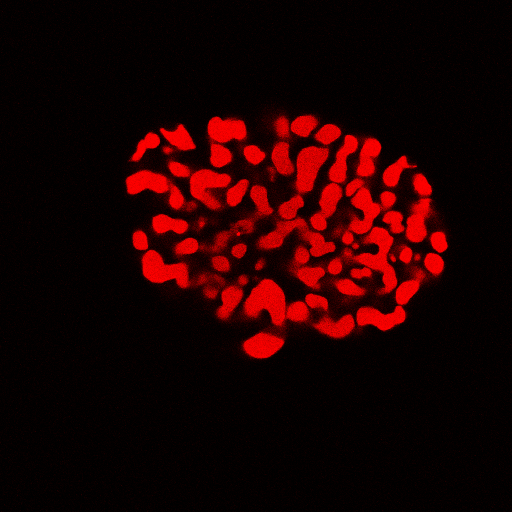
single orthoslice through glomerulus cast |
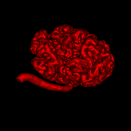
rendered glomerulus model |
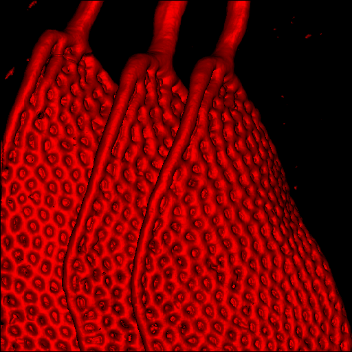
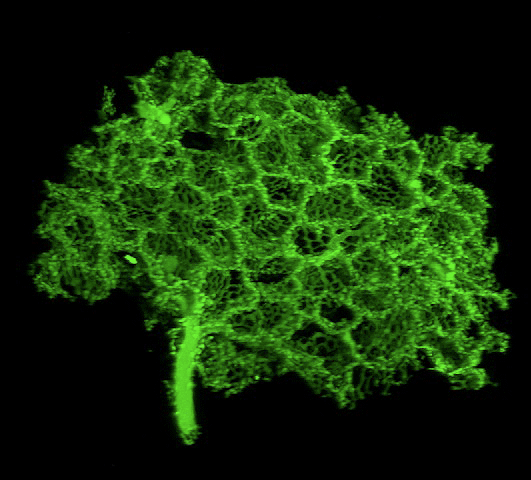
rendered fish gills model |
 |
 |
 |
 |
model of choriocapillaris capillaries of the eye |
model of retinal capillaries |
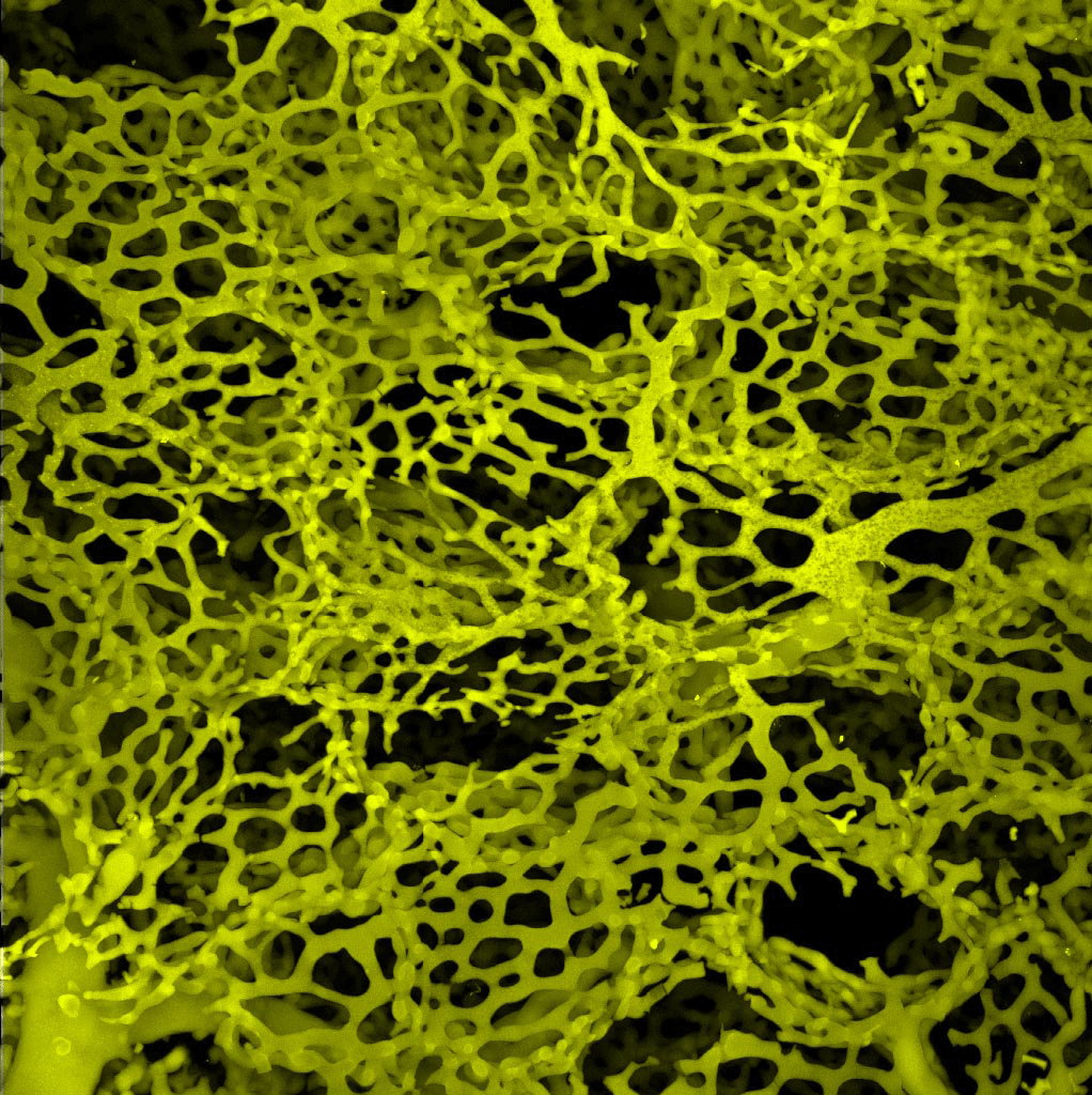
projection view of lung capillaries model |
model of alveolar capillaries of lung |
 |
 |
 |
 |
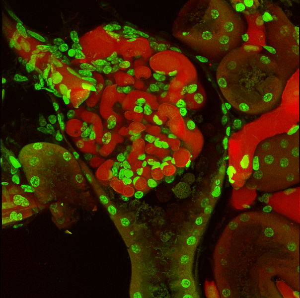
uncorroded glomerulus and Bowman's capsule |
uncorroded glomerulus and arteriole |
uncorroded glomeruli and kidney tubules |
uncorroded fish gill capillaries |