Three Dimensional Models
of Tissue and Cell Structure
Click on button next to image title to retrieve image:
axoneme (9+2)
kidney nephron
muscle sarcomere
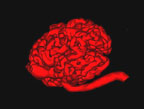
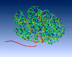
capillary tuft
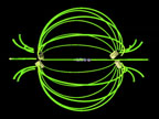
glomerular capillaries #1
small vein & tunics
ciliated cell composite
glomerular capillaries #2
smooth muscle fiber
ciliated cell
goblet cells
spinal cord model
columnar epithelium
lumpy cell
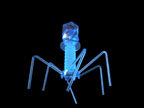
T4 phage model
squamous epithelium
membrane bilayer model
thyroid follicles
epithelium model #1
tubulin monomers
Connexons
epithelium model #2
renal corpuscle
Axon
epithelium model #3
rod cell
Connective Tissue
erythrocytes
rod and cone cell
Eosinophil
freeze-fracture model
salivon
Spermatazoan
Epidermis
Smooth Muscle Fiber 1
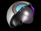
Eye
Glomerular Capillaries
Smooth Muscle Fibers
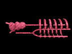
Skeletal Muscle Fiber
Unmyelinated Axons
Animated Models of Tissues and Cell Structure
Eye Model
Aortic Arches
Spindle Apparatus
Nephron Model
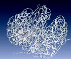
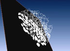
Glomerular Corrosion Cast
Skeletal Model of Corrosion Cast
Color-Coded Skeleton
Orthoslice through Skeleton
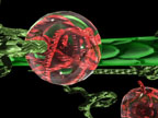
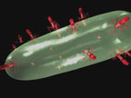
Bacterium w Phages
Phage Docking
Ovary
Bacterial Fission
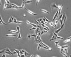
Cultured Cells
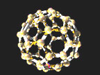
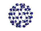
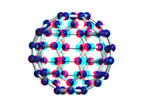
Buckyball Flythrough
Buckyball
Buckyball Anaglyph